NMR structure of a parallel-stranded DNA duplex at atomic resolution.
Parvathy, V.R., Bhaumik, S.R., Chary, K.V., Govil, G., Liu, K., Howard, F.B., Miles, H.T.(2002) Nucleic Acids Res 30: 1500-1511
- PubMed: 11917010
- DOI: https://doi.org/10.1093/nar/30.7.1500
- Primary Citation of Related Structures:
1JUU - PubMed Abstract:
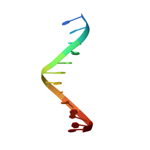
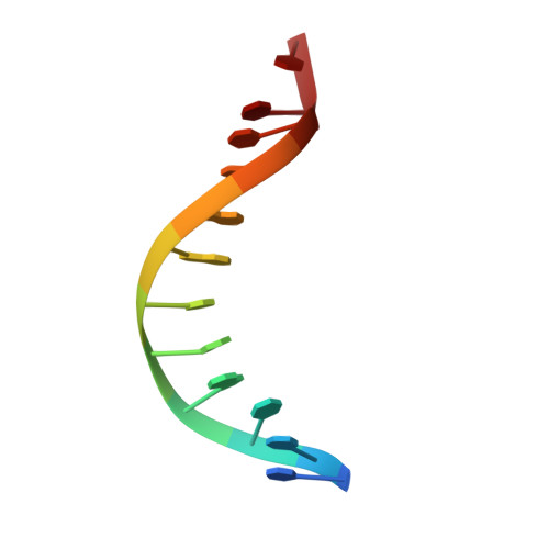
DNA dodecamers have been designed with two cytosines on each end and intervening A and T stretches, such that the oligomers have fully complementary A:T base pairs when aligned in the parallel orientation. Spectroscopic (UV, CD and IR), NMR and molecular dynamics studies have shown that oligomers having the sequences d(CCATAATTTACC) and d(CCTATTAAATCC) form a parallel-stranded duplex when dissolved at 1:1 stoichiometry in aqueous solution. This is due to the C:C+ clamps on either end and extensive mismatches in the antiparallel orientation. The structure is stable at neutral and acidic pH. At higher temperatures, the duplex melts into single strands in a highly cooperative fashion. All adenine, cytosine and thymine nucleotides adopt the anti conformation with respect to the glycosidic bond. The A:T base pairs form reverse Watson-Crick base pairs. The duplex shows base stacking and NOEs between the base protons T(H6)/A(H8) and the sugar protons (H1'/H2'/H2") of the preceding nucleotide, as has been observed in antiparallel duplexes. However, no NOEs are observed between base protons H2/H6/H8 of sequential nucleotides, though such NOEs are observed between T(CH3) and A(H8). A three-dimensional structure of the parallel-stranded duplex at atomic resolution has been obtained using molecular dynamics simulations under NMR constraints. The simulated structures have torsional angles very similar to those found in B-DNA duplexes, but the base stacking and helicoid parameters are significantly different.
Organizational Affiliation:
Department of Chemical Sciences, Tata Institute of Fundamental Research, Homi Bhabha Road, Colaba, Mumbai 400 005, India.