1JPW

Crystal Structure of a Human Tcf-4 / beta-Catenin Complex


Experimental Data Snapshot

  • Method: X-RAY DIFFRACTION
  • Resolution: 2.50 Å
  • R-Value Free: 0.390 
  • R-Value Work: 0.304 
  • R-Value Observed: 0.304 

wwPDB Validation   3D Report Full Report


This is version 1.4 of the entry. See complete history


Literature

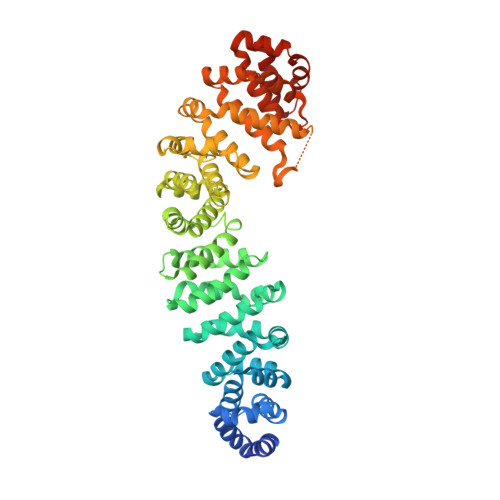
Structure of a human Tcf4-beta-catenin complex.

Poy, F.Lepourcelet, M.Shivdasani, R.A.Eck, M.J.

(2001) Nat Struct Biol 8: 1053-1057

  • DOI: https://doi.org/10.1038/nsb720
  • Primary Citation of Related Structures:  
    1JPW

  • PubMed Abstract: 

    The multifunctional protein beta-catenin is important for cell adhesion, because it binds cadherins, and the Wnt signal transduction pathway, where it interacts with the Adenomatous polyposis coli (APC) protein and TCF/Lef family transcription factors. Mutations in APC or in beta-catenin are estimated to trigger formation of over 90% of all colon cancers. In colonic epithelia, these mutations produce elevated levels of Tcf4-beta-catenin, which stimulates a transcriptional response that initiates polyp formation and eventually malignant growth. Thus, disruption of the Tcf4-beta-catenin interaction may be an attractive goal for therapeutic intervention. Here we describe the crystal structure of a human Tcf4-beta-catenin complex and compare it with recent structures of beta-catenin in complex with Xenopus Tcf3 (XTcf3) and mammalian E-cadherin. The structure reveals anticipated similarities with the closely related XTcf3 complex but unexpectedly lacks one component observed in the XTcf3 structure.


  • Organizational Affiliation

    Department of Cancer Biology, Dana-Farber Cancer Institute, 44 Binney Street, Boston, Massachusetts 02115, USA.


Macromolecules
Find similar proteins by:  (by identity cutoff)  |  3D Structure
Entity ID: 1
MoleculeChains Sequence LengthOrganismDetailsImage
BETA-CATENINA,
C [auth B],
E [auth C]
540Homo sapiensMutation(s): 0 
UniProt & NIH Common Fund Data Resources
Find proteins for P35222 (Homo sapiens)
Explore P35222 
Go to UniProtKB:  P35222
PHAROS:  P35222
GTEx:  ENSG00000168036 
Entity Groups  
Sequence Clusters30% Identity50% Identity70% Identity90% Identity95% Identity100% Identity
UniProt GroupP35222
Sequence Annotations
Expand
  • Reference Sequence
Find similar proteins by:  (by identity cutoff)  |  3D Structure
Entity ID: 2
MoleculeChains Sequence LengthOrganismDetailsImage
transcription factor 7-like 2B [auth D],
D [auth E],
F
49Homo sapiensMutation(s): 0 
UniProt & NIH Common Fund Data Resources
Find proteins for Q9NQB0 (Homo sapiens)
Explore Q9NQB0 
Go to UniProtKB:  Q9NQB0
PHAROS:  Q9NQB0
GTEx:  ENSG00000148737 
Entity Groups  
Sequence Clusters30% Identity50% Identity70% Identity90% Identity95% Identity100% Identity
UniProt GroupQ9NQB0
Sequence Annotations
Expand
  • Reference Sequence
Experimental Data & Validation

Experimental Data

  • Method: X-RAY DIFFRACTION
  • Resolution: 2.50 Å
  • R-Value Free: 0.390 
  • R-Value Work: 0.304 
  • R-Value Observed: 0.304 
  • Space Group: C 1 2 1
Unit Cell:
Length ( Å )Angle ( ˚ )
a = 376.53α = 90
b = 92.65β = 95.98
c = 49.25γ = 90
Software Package:
Software NamePurpose
CNSrefinement
DENZOdata reduction
SCALEPACKdata scaling
CNSphasing

Structure Validation

View Full Validation Report


Entry History 

Deposition Data

Revision History  (Full details and data files)

  • Version 1.0: 2001-12-05
    Type: Initial release
  • Version 1.1: 2008-04-27
    Changes: Version format compliance
  • Version 1.2: 2011-07-13
    Changes: Version format compliance
  • Version 1.3: 2018-01-31
    Changes: Experimental preparation
  • Version 1.4: 2023-08-16
    Changes: Data collection, Database references, Refinement description