1JLM

I-DOMAIN FROM INTEGRIN CR3, MN2+ BOUND


Experimental Data Snapshot

  • Method: X-RAY DIFFRACTION
  • Resolution: 2.00 Å
  • R-Value Work: 0.219 
  • R-Value Observed: 0.219 

wwPDB Validation   3D Report Full Report


This is version 1.4 of the entry. See complete history


Literature

Two conformations of the integrin A-domain (I-domain): a pathway for activation?

Lee, J.O.Bankston, L.A.Arnaout, M.A.Liddington, R.C.

(1995) Structure 3: 1333-1340

  • DOI: https://doi.org/10.1016/s0969-2126(01)00271-4
  • Primary Citation of Related Structures:  
    1JLM

  • PubMed Abstract: 

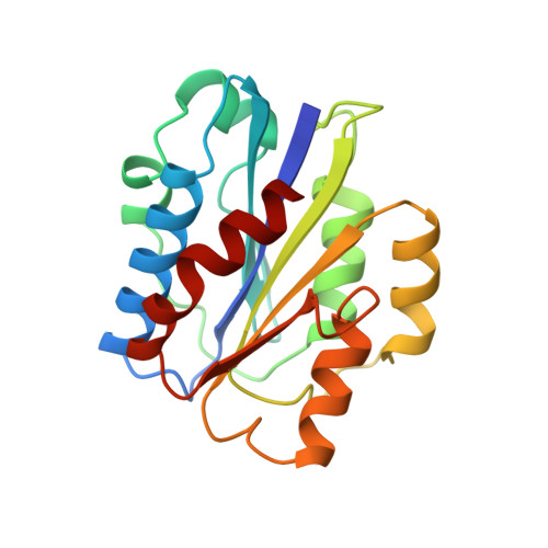
    Integrins are plasma membrane proteins that mediate adhesion to other cells and to components of the extracellular matrix. Most integrins are constitutively inactive in resting cells, but are rapidly and reversibly activated in response to agonists, leading to highly regulated cell adhesion. This activation is associated with conformational changes in their extracellular portions, but the nature of the structural changes that lead to a change in adhesiveness is not understood. The interactions of several integrins with their extracellular ligands are mediated by an A-type domain (generally called the I-domain in integrins). Binding of the I-domain to protein ligands is dependent on divalent cations. We have described previously the structure of the I-domain from complement receptor 3 with bound Mg2+, in which the glutamate side chain from a second I-domain completes the octahedral coordination sphere of the metal, acting as a ligand mimetic.


  • Organizational Affiliation

    Dana-Farber Cancer Institute, Harvard Medical School, Boston, MA 02115, USA.


Macromolecules
Find similar proteins by:  (by identity cutoff)  |  3D Structure
Entity ID: 1
MoleculeChains Sequence LengthOrganismDetailsImage
INTEGRIN192Homo sapiensMutation(s): 0 
UniProt & NIH Common Fund Data Resources
Find proteins for P11215 (Homo sapiens)
Explore P11215 
Go to UniProtKB:  P11215
PHAROS:  P11215
GTEx:  ENSG00000169896 
Entity Groups  
Sequence Clusters30% Identity50% Identity70% Identity90% Identity95% Identity100% Identity
UniProt GroupP11215
Sequence Annotations
Expand
  • Reference Sequence
Small Molecules
Ligands 1 Unique
IDChains Name / Formula / InChI Key2D Diagram3D Interactions
MN
Query on MN

Download Ideal Coordinates CCD File 
B [auth A]MANGANESE (II) ION
Mn
WAEMQWOKJMHJLA-UHFFFAOYSA-N
Experimental Data & Validation

Experimental Data

  • Method: X-RAY DIFFRACTION
  • Resolution: 2.00 Å
  • R-Value Work: 0.219 
  • R-Value Observed: 0.219 
  • Space Group: C 1 2 1
Unit Cell:
Length ( Å )Angle ( ˚ )
a = 135α = 90
b = 37.1β = 92.6
c = 38.5γ = 90
Software Package:
Software NamePurpose
X-PLORmodel building
X-PLORrefinement
DENZOdata reduction
SCALEPACKdata scaling
X-PLORphasing

Structure Validation

View Full Validation Report


Entry History 

Deposition Data

Revision History  (Full details and data files)

  • Version 1.0: 1997-01-11
    Type: Initial release
  • Version 1.1: 2008-03-24
    Changes: Version format compliance
  • Version 1.2: 2011-07-13
    Changes: Version format compliance
  • Version 1.3: 2018-03-21
    Changes: Data collection, Other
  • Version 1.4: 2023-08-09
    Changes: Database references, Derived calculations, Refinement description