Inhibition mechanism of cytokine activity of human autocrine motility factor examined by crystal structure analyses and site-directed mutagenesis studies.
Tanaka, N., Haga, A., Uemura, H., Akiyama, H., Funasaka, T., Nagase, H., Raz, A., Nakamura, K.T.(2002) J Mol Biol 318: 985-997
- PubMed: 12054796
- DOI: https://doi.org/10.1016/S0022-2836(02)00186-9
- Primary Citation of Related Structures:
1IRI, 1JIQ - PubMed Abstract:
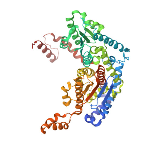
Autocrine motility factor (AMF), a tumor-secreted cytokine, stimulates cell migration in vitro and metastasis in vivo. AMF is genetically identical with the extracellular cytokines neuroleukin (NLK) and maturation factor (MF) and, interestingly, the intracellular enzyme phosphohexose isomerase (PHI). The crystal structures of the inhibitor-free open form and the inhibitor (erythrose 4-phosphate, E4P, a strong inhibitor of AMF's cytokine activity)-bound closed form of human AMF have been determined at 1.9 A and 2.4 A resolution, respectively. Upon E4P binding, local conformation changes (open to closed) occur around the inhibitor-binding site. The E4P-bound structure shows that the location of the inhibitor (of cytokine activity) binding site of human AMF is very similar to those of the inhibitor (of enzymatic activity) binding sites of PHIs. The present study shows clearly that there is structural overlap of the regions responsible for the enzymatic and cytokine functions of AMF and PHI and suggests two scenarios for the inhibition mechanism of cytokine activity of AMF by the carbohydrate phosphate group. One likely scenario is that the compound could compete for AMF binding with the carbohydrate moiety of the AMF receptor (AMFR), which is a glycosylated seven-transmembrane helix protein. The other scenario is that the local conformation changes upon inhibitor binding may affect the AMF-AMFR interactions. To examine roles of the residues in the inhibitor-binding site, two mutant AMFs were prepared. Replacements of His389, which is hydrogen-bonded to the hydroxyl group of E4P by Phe, and Thr215, which is hydrogen-bonded to the phosphate group of E4P by Asp, result in mutant AMFs that are impaired in cytokine activity. These results suggest a role for these amino acids in recognition of a carbohydrate moiety of the AMFR. Since the E4P is one of the smallest compounds having AMF inhibitor activity, knowledge of the present crystal structure would provide an insight into the lead compound design of more effective AMF inhibitors.
Organizational Affiliation:
School of Pharmaceutical Sciences, Showa University, 1-5-8 Hatanodai, Shinagawa-ku, Tokyo 142-8555, Japan.