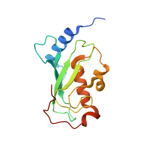
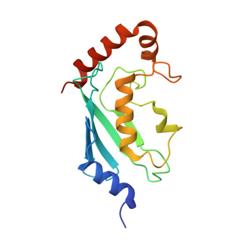
1J7D

Crystal Structure of hMms2-hUbc13


Experimental Data Snapshot

  • Method: X-RAY DIFFRACTION
  • Resolution: 1.85 Å
  • R-Value Free: 0.244 
  • R-Value Work: 0.215 

wwPDB Validation   3D Report Full Report


This is version 1.3 of the entry. See complete history


Literature

Crystal structure of the human ubiquitin conjugating enzyme complex, hMms2-hUbc13.

Moraes, T.F.Edwards, R.A.McKenna, S.Pastushok, L.Xiao, W.Glover, J.N.Ellison, M.J.

(2001) Nat Struct Biol 8: 669-673

  • DOI: https://doi.org/10.1038/90373
  • Primary Citation of Related Structures:  
    1J74, 1J7D

  • PubMed Abstract: 

    The ubiquitin conjugating enzyme complex Mms2-Ubc13 plays a key role in post-replicative DNA repair in yeast and the NF-kappaB signal transduction pathway in humans. This complex assembles novel polyubiquitin chains onto yet uncharacterized protein targets. Here we report the crystal structure of a complex between hMms2 (Uev1) and hUbc13 at 1.85 A resolution and a structure of free hMms2 at 1.9 A resolution. These structures reveal that the hMms2 monomer undergoes a localized conformational change upon interaction with hUbc13. The nature of the interface provides a physical basis for the preference of Mms2 for Ubc13 as a partner over a variety of other structurally similar ubiquitin-conjugating enzymes. The structure of the hMms2-hUbc13 complex provides the conceptual foundation for understanding the mechanism of Lys 63 multiubiquitin chain assembly and for its interactions with the RING finger proteins Rad5 and Traf6.


  • Organizational Affiliation

    Department of Biochemistry, University of Alberta, Edmonton, Alberta, T6G-2H7, Canada.


Macromolecules
Find similar proteins by:  (by identity cutoff)  |  3D Structure
Entity ID: 1
MoleculeChains Sequence LengthOrganismDetailsImage
MMS2145Homo sapiensMutation(s): 0 
UniProt & NIH Common Fund Data Resources
Find proteins for Q15819 (Homo sapiens)
Explore Q15819 
Go to UniProtKB:  Q15819
PHAROS:  Q15819
GTEx:  ENSG00000169139 
Entity Groups  
Sequence Clusters30% Identity50% Identity70% Identity90% Identity95% Identity100% Identity
UniProt GroupQ15819
Sequence Annotations
Expand
  • Reference Sequence
Find similar proteins by:  (by identity cutoff)  |  3D Structure
Entity ID: 2
MoleculeChains Sequence LengthOrganismDetailsImage
UBIQUITIN-CONJUGATING ENZYME E2-17 KDA152Homo sapiensMutation(s): 0 
EC: 6.3.2.19
UniProt & NIH Common Fund Data Resources
Find proteins for P61088 (Homo sapiens)
Explore P61088 
Go to UniProtKB:  P61088
PHAROS:  P61088
GTEx:  ENSG00000177889 
Entity Groups  
Sequence Clusters30% Identity50% Identity70% Identity90% Identity95% Identity100% Identity
UniProt GroupP61088
Sequence Annotations
Expand
  • Reference Sequence
Experimental Data & Validation

Experimental Data

  • Method: X-RAY DIFFRACTION
  • Resolution: 1.85 Å
  • R-Value Free: 0.244 
  • R-Value Work: 0.215 
  • Space Group: P 21 21 21
Unit Cell:
Length ( Å )Angle ( ˚ )
a = 44.132α = 90
b = 74.137β = 90
c = 91.489γ = 90
Software Package:
Software NamePurpose
DENZOdata reduction
SCALEPACKdata scaling
SOLVEphasing
CNSrefinement

Structure Validation

View Full Validation Report


Entry History 

Deposition Data

Revision History  (Full details and data files)

  • Version 1.0: 2001-08-08
    Type: Initial release
  • Version 1.1: 2008-04-27
    Changes: Version format compliance
  • Version 1.2: 2011-07-13
    Changes: Version format compliance
  • Version 1.3: 2024-02-07
    Changes: Data collection, Database references