Crystal structures of recombinant and native soybean beta-conglycinin beta homotrimers.
Maruyama, N., Adachi, M., Takahashi, K., Yagasaki, K., Kohno, M., Takenaka, Y., Okuda, E., Nakagawa, S., Mikami, B., Utsumi, S.(2001) Eur J Biochem 268: 3595-3604
- PubMed: 11422391
- DOI: https://doi.org/10.1046/j.1432-1327.2001.02268.x
- Primary Citation of Related Structures:
1IPK - PubMed Abstract:
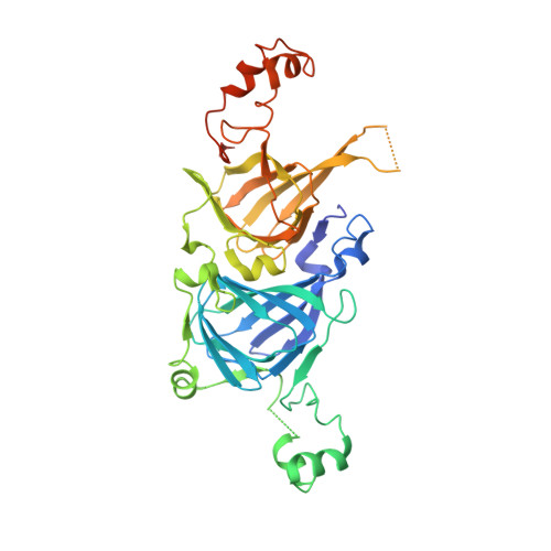
The crystal structures of recombinant and native beta homotrimers of soybean beta-conglycinin were determined by X-ray crystallography at 2.7 and 2.8 A resolutions, respectively. The crystals of the recombinant and native beta homotrimers belong to space group P21 with cell parameters a = 80.51 A, b = 63.48 A, c = 131.43 A, and beta = 90.01 degrees and with cell parameters a = 82.78 A, b = 69.47 A, c = 125.33 A and beta = 97.22 degrees, respectively. The beta monomers consist of amino-terminal and carboxyl-terminal modules that are very similar to each other and are related by a pseudo-dyad axis. Each module of the beta monomer is subdivided into a core and a loop domain. These structural features of both beta homotrimers are consistent with those of canavalin and phaseolin, which are similar vicilin class proteins. The superposition of the models of the native and recombinant beta monomers shows a root mean square deviation of 0.43-0.51 A for 343 common Calpha atoms within 2.0 A. This result indicates that the N-linked glycans do not influence the final structure of the beta homotrimer. Comparison of the models of beta-conglycinin, phaseolin and canavalin indicates that beta-conglycinin resembles canavalin rather than phaseolin, and that canavalin and phaseolin differ the most among them. The evolutional relationships are discussed.
Organizational Affiliation:
Research Institute for Food Science, Kyoto University, Uji, Japan.