1IOD

CRYSTAL STRUCTURE OF THE COMPLEX BETWEEN THE COAGULATION FACTOR X BINDING PROTEIN FROM SNAKE VENOM AND THE GLA DOMAIN OF FACTOR X


Experimental Data Snapshot

  • Method: X-RAY DIFFRACTION
  • Resolution: 2.30 Å
  • R-Value Free: 0.267 
  • R-Value Work: 0.201 
  • R-Value Observed: 0.201 

wwPDB Validation   3D Report Full Report


This is version 1.4 of the entry. See complete history


Literature

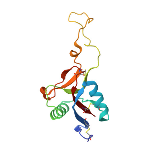
Crystal structure of an anticoagulant protein in complex with the Gla domain of factor X.

Mizuno, H.Fujimoto, Z.Atoda, H.Morita, T.

(2001) Proc Natl Acad Sci U S A 98: 7230-7234

  • DOI: https://doi.org/10.1073/pnas.131179698
  • Primary Citation of Related Structures:  
    1IOD

  • PubMed Abstract: 

    The gamma-carboxyglutamic acid (Gla) domain of blood coagulation factors is responsible for Ca2+-dependent phospholipid membrane binding. Factor X-binding protein (X-bp), an anticoagulant protein from snake venom, specifically binds to the Gla domain of factor X. The crystal structure of X-bp in complex with the Gla domain peptide of factor X at 2.3-A resolution showed that the anticoagulation is based on the fact that two patches of the Gla domain essential for membrane binding are buried in the complex formation. The Gla domain thus is expected to be a new target of anticoagulant drugs, and X-bp provides a basis for designing them. This structure also provides a membrane-bound model of factor X.


  • Organizational Affiliation

    Department of Biotechnology, National Institute of Agrobiological Resources, Tsukuba, Ibaraki 305-8602, Japan. mizuno@nias.affrc.go.jp


Macromolecules
Find similar proteins by:  (by identity cutoff)  |  3D Structure
Entity ID: 1
MoleculeChains Sequence LengthOrganismDetailsImage
COAGULATION FACTOR X BINDING PROTEIN129Deinagkistrodon acutusMutation(s): 0 
Membrane Entity: Yes 
UniProt
Find proteins for Q9IAM1 (Deinagkistrodon acutus)
Explore Q9IAM1 
Go to UniProtKB:  Q9IAM1
Entity Groups  
Sequence Clusters30% Identity50% Identity70% Identity90% Identity95% Identity100% Identity
UniProt GroupQ9IAM1
Sequence Annotations
Expand
  • Reference Sequence
Find similar proteins by:  (by identity cutoff)  |  3D Structure
Entity ID: 2
MoleculeChains Sequence LengthOrganismDetailsImage
COAGULATION FACTOR X BINDING PROTEIN123Deinagkistrodon acutusMutation(s): 0 
Membrane Entity: Yes 
UniProt
Find proteins for Q9DEF8 (Deinagkistrodon acutus)
Explore Q9DEF8 
Go to UniProtKB:  Q9DEF8
Entity Groups  
Sequence Clusters30% Identity50% Identity70% Identity90% Identity95% Identity100% Identity
UniProt GroupQ9DEF8
Sequence Annotations
Expand
  • Reference Sequence
Find similar proteins by:  (by identity cutoff)  |  3D Structure
Entity ID: 3
MoleculeChains Sequence LengthOrganismDetailsImage
COAGULATION FACTOR X GLA DOMAINC [auth G]44Bos taurusMutation(s): 0 
EC: 3.4.21.6
Membrane Entity: Yes 
UniProt
Find proteins for P00743 (Bos taurus)
Explore P00743 
Go to UniProtKB:  P00743
Entity Groups  
Sequence Clusters30% Identity50% Identity70% Identity90% Identity95% Identity100% Identity
UniProt GroupP00743
Sequence Annotations
Expand
  • Reference Sequence
Small Molecules
Ligands 1 Unique
IDChains Name / Formula / InChI Key2D Diagram3D Interactions
CA
Query on CA

Download Ideal Coordinates CCD File 
D [auth A]
E [auth A]
F [auth A]
G [auth B]
H [auth G]
D [auth A],
E [auth A],
F [auth A],
G [auth B],
H [auth G],
I [auth G],
J [auth G],
K [auth G],
L [auth G],
M [auth G],
N [auth G],
O [auth G]
CALCIUM ION
Ca
BHPQYMZQTOCNFJ-UHFFFAOYSA-N
Modified Residues  1 Unique
IDChains TypeFormula2D DiagramParent
CGU
Query on CGU
C [auth G]L-PEPTIDE LINKINGC6 H9 N O6GLU
Experimental Data & Validation

Experimental Data

  • Method: X-RAY DIFFRACTION
  • Resolution: 2.30 Å
  • R-Value Free: 0.267 
  • R-Value Work: 0.201 
  • R-Value Observed: 0.201 
  • Space Group: P 41 21 2
Unit Cell:
Length ( Å )Angle ( ˚ )
a = 99.76α = 90
b = 99.76β = 90
c = 90.42γ = 90
Software Package:
Software NamePurpose
DENZOdata reduction
SCALEPACKdata scaling
AMoREphasing
X-PLORrefinement

Structure Validation

View Full Validation Report


Entry History 

Deposition Data

Revision History  (Full details and data files)

  • Version 1.0: 2001-06-27
    Type: Initial release
  • Version 1.1: 2008-04-27
    Changes: Version format compliance
  • Version 1.2: 2011-07-13
    Changes: Version format compliance
  • Version 1.3: 2023-12-27
    Changes: Data collection, Database references, Derived calculations
  • Version 1.4: 2024-04-03
    Changes: Refinement description