1IO0

CRYSTAL STRUCTURE OF TROPOMODULIN C-TERMINAL HALF


Experimental Data Snapshot

  • Method: X-RAY DIFFRACTION
  • Resolution: 1.45 Å
  • R-Value Free: 0.220 
  • R-Value Work: 0.203 

wwPDB Validation   3D Report Full Report


This is version 1.4 of the entry. See complete history


Literature

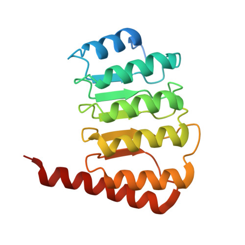
Crystal structure of the C-terminal half of tropomodulin and structural basis of actin filament pointed-end capping.

Krieger, I.Kostyukova, A.Yamashita, A.Nitanai, Y.Maeda, Y.

(2002) Biophys J 83: 2716-2725

  • DOI: https://doi.org/10.1016/S0006-3495(02)75281-8
  • Primary Citation of Related Structures:  
    1IO0

  • PubMed Abstract: 

    Tropomodulin is the unique pointed-end capping protein of the actin-tropomyosin filament. By blocking elongation and depolymerization, tropomodulin regulates the architecture and the dynamics of the filament. Here we report the crystal structure at 1.45-A resolution of the C-terminal half of tropomodulin (C20), the actin-binding moiety of tropomodulin. C20 is a leucine-rich repeat domain, and this is the first actin-associated protein with a leucine-rich repeat. Binding assays suggested that C20 also interacts with the N-terminal fragment, M1-M2-M3, of nebulin. Based on the crystal structure, we propose a model for C20 docking to the actin subunit at the pointed end. Although speculative, the model is consistent with the idea that a tropomodulin molecule competes with an actin subunit for a pointed end. The model also suggests that interactions with tropomyosin, actin, and nebulin are all possible sources of influences on the dynamic properties of pointed-end capping by tropomodulin.


  • Organizational Affiliation

    Laboratory for Structural Biochemistry, RIKEN Harima Institute at SPring-8, Mikazuki, Sayo, Hyogo, Japan 679-5148.


Macromolecules
Find similar proteins by:  (by identity cutoff)  |  3D Structure
Entity ID: 1
MoleculeChains Sequence LengthOrganismDetailsImage
TROPOMODULIN185Gallus gallusMutation(s): 0 
UniProt
Find proteins for Q9DEA6 (Gallus gallus)
Explore Q9DEA6 
Go to UniProtKB:  Q9DEA6
Entity Groups  
Sequence Clusters30% Identity50% Identity70% Identity90% Identity95% Identity100% Identity
UniProt GroupQ9DEA6
Sequence Annotations
Expand
  • Reference Sequence
Small Molecules
Ligands 1 Unique
IDChains Name / Formula / InChI Key2D Diagram3D Interactions
ZN
Query on ZN

Download Ideal Coordinates CCD File 
B [auth A]ZINC ION
Zn
PTFCDOFLOPIGGS-UHFFFAOYSA-N
Experimental Data & Validation

Experimental Data

  • Method: X-RAY DIFFRACTION
  • Resolution: 1.45 Å
  • R-Value Free: 0.220 
  • R-Value Work: 0.203 
  • Space Group: H 3
Unit Cell:
Length ( Å )Angle ( ˚ )
a = 69.294α = 90
b = 69.294β = 90
c = 101.223γ = 120
Software Package:
Software NamePurpose
DENZOdata reduction
SCALEPACKdata scaling
SOLVEphasing
DMmodel building
ARP/wARPmodel building
X-PLORrefinement
DMphasing

Structure Validation

View Full Validation Report


Entry History 

Deposition Data

Revision History  (Full details and data files)

  • Version 1.0: 2002-11-27
    Type: Initial release
  • Version 1.1: 2008-04-27
    Changes: Version format compliance
  • Version 1.2: 2011-07-13
    Changes: Version format compliance
  • Version 1.3: 2019-10-23
    Changes: Data collection, Database references
  • Version 1.4: 2023-12-27
    Changes: Data collection, Database references, Derived calculations