1I3T

MOLECULAR AND CRYSTAL STRUCTURE OF D(CGCGAATT(MO4)CGCG): THE WATSON-CRICK TYPE AND WOBBLE N4-METHOXYCYTIDINE/GUANOSINE BASE PAIRS IN B-DNA


Experimental Data Snapshot

  • Method: X-RAY DIFFRACTION
  • Resolution: 1.60 Å
  • R-Value Free: 0.256 
  • R-Value Work: 0.224 

wwPDB Validation   3D Report Full Report


This is version 1.3 of the entry. See complete history


Literature

Crystallographic studies on damaged DNAs: III. N(4)-methoxycytosine can form both Watson-Crick type and wobbled base pairs in a B-form duplex.

Hossain, M.T.Chatake, T.Hikima, T.Tsunoda, M.Sunami, T.Ueno, Y.Matsuda, A.Takenaka, A.

(2001) J Biochem 130: 9-12

  • DOI: https://doi.org/10.1093/oxfordjournals.jbchem.a002967
  • Primary Citation of Related Structures:  
    1I3T, 1I47

  • PubMed Abstract: 

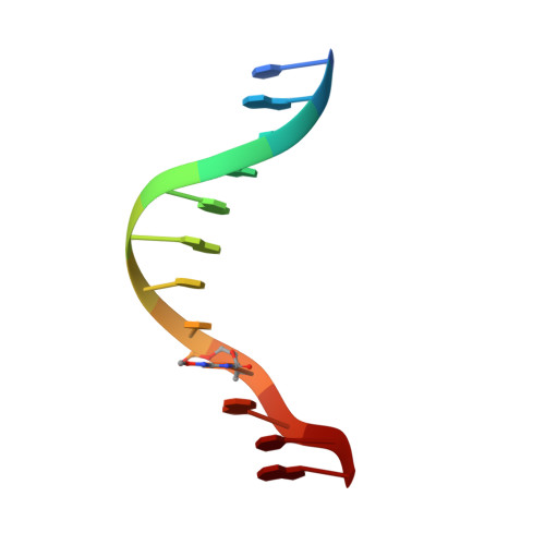
    To investigate the mutation mechanism of purine transition in DNA damaged with methoxyamine, a DNA dodecamer with the sequence d(CGCGAATTmo(4)CGCG), where mo(4)C is 2'-deoxy-N(4)-methoxycytidine, has been synthesized and its crystal structure determined. Two dodecamers form a B-form duplex. Electron density maps clearly show that one of the two mo(4)C residues forms a pair with a guanine residue of the opposite strand, the geometry being the canonical Watson-Crick type, and that the other mo(4)C residue forms a wobble pair with the opposite guanine residue. These two pairings are ascribed to the tautomerization of the methoxylated cytosine moieties between the amino and imino forms.


  • Organizational Affiliation

    Graduate School of Bioscience and Biotechnology, Tokyo Institute of Technology, Nagatsuta, Midori-ku, Yokohama 226-8501, Japan.


Macromolecules

Find similar nucleic acids by:  Sequence   |   3D Structure  

Entity ID: 1
MoleculeChains LengthOrganismImage
5'-D(*CP*GP*CP*GP*AP*AP*TP*TP*(C45)P*GP*CP*GP)-3'
A, B
12N/A
Sequence Annotations
Expand
  • Reference Sequence
Small Molecules
Ligands 1 Unique
IDChains Name / Formula / InChI Key2D Diagram3D Interactions
MG
Query on MG

Download Ideal Coordinates CCD File 
C [auth A]MAGNESIUM ION
Mg
JLVVSXFLKOJNIY-UHFFFAOYSA-N
Experimental Data & Validation

Experimental Data

  • Method: X-RAY DIFFRACTION
  • Resolution: 1.60 Å
  • R-Value Free: 0.256 
  • R-Value Work: 0.224 
  • Space Group: P 21 21 21
Unit Cell:
Length ( Å )Angle ( ˚ )
a = 25.182α = 90
b = 40.877β = 90
c = 63.731γ = 90
Software Package:
Software NamePurpose
AMoREphasing
CNSrefinement
DENZOdata reduction
SCALEPACKdata scaling

Structure Validation

View Full Validation Report


Entry History 

Deposition Data

Revision History  (Full details and data files)

  • Version 1.0: 2001-09-14
    Type: Initial release
  • Version 1.1: 2007-10-16
    Changes: Version format compliance
  • Version 1.2: 2011-07-13
    Changes: Version format compliance
  • Version 1.3: 2023-08-09
    Changes: Data collection, Database references, Derived calculations, Refinement description