1I0E

CRYSTAL STRUCTURE OF CREATINE KINASE FROM HUMAN MUSCLE


Experimental Data Snapshot

  • Method: X-RAY DIFFRACTION
  • Resolution: 3.50 Å
  • R-Value Free: 0.287 
  • R-Value Work: 0.211 
  • R-Value Observed: 0.211 

wwPDB Validation   3D Report Full Report


This is version 1.5 of the entry. See complete history


Literature

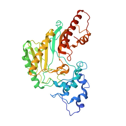
Structure of human muscle creatine kinase.

Shen, Y.Q.Tang, L.Zhou, H.M.Lin, Z.J.

(2001) Acta Crystallogr D Biol Crystallogr 57: 1196-1200

  • DOI: https://doi.org/10.1107/s0907444901007703
  • Primary Citation of Related Structures:  
    1I0E

  • PubMed Abstract: 

    The crystal structure of human muscle creatine kinase has been determined by the molecular-replacement method and refined at 3.5A resolution. The structures of both the monomer and the dimer closely resemble those of the other known structures in the creatine kinase family. Two types of dimers, one with a non-crystallographic twofold symmetry axis and the other with a crystallographic twofold symmetry axis, were found to exist simultaneously in the crystal. These dimers form an infinite "double-helix"-like structure along an unusual long crystallographic 3(1) axis.


  • Organizational Affiliation

    National Laboratory of Biological Macromolecules, Institute of Biophysics, Academica Sinica, Beijing 100101, People's Republic of China.


Macromolecules
Find similar proteins by:  (by identity cutoff)  |  3D Structure
Entity ID: 1
MoleculeChains Sequence LengthOrganismDetailsImage
CREATINE KINASE,M CHAIN
A, B, C, D
381Homo sapiensMutation(s): 0 
EC: 2.7.3.2
UniProt & NIH Common Fund Data Resources
Find proteins for P06732 (Homo sapiens)
Explore P06732 
Go to UniProtKB:  P06732
PHAROS:  P06732
GTEx:  ENSG00000104879 
Entity Groups  
Sequence Clusters30% Identity50% Identity70% Identity90% Identity95% Identity100% Identity
UniProt GroupP06732
Sequence Annotations
Expand
  • Reference Sequence
Experimental Data & Validation

Experimental Data

  • Method: X-RAY DIFFRACTION
  • Resolution: 3.50 Å
  • R-Value Free: 0.287 
  • R-Value Work: 0.211 
  • R-Value Observed: 0.211 
  • Space Group: P 31 1 2
Unit Cell:
Length ( Å )Angle ( ˚ )
a = 89.326α = 90
b = 89.326β = 90
c = 402.962γ = 120
Software Package:
Software NamePurpose
DENZOdata reduction
SCALEPACKdata scaling
AMoREphasing
CNSrefinement

Structure Validation

View Full Validation Report


Entry History 

Deposition Data

Revision History  (Full details and data files)

  • Version 1.0: 2003-04-01
    Type: Initial release
  • Version 1.1: 2008-04-27
    Changes: Version format compliance
  • Version 1.2: 2011-07-13
    Changes: Version format compliance
  • Version 1.3: 2019-10-23
    Changes: Data collection, Database references
  • Version 1.4: 2024-03-13
    Changes: Data collection, Database references
  • Version 1.5: 2024-04-03
    Changes: Refinement description