1HQP

CRYSTAL STRUCTURE OF A TRUNCATED FORM OF PORCINE ODORANT-BINDING PROTEIN


Experimental Data Snapshot

  • Method: X-RAY DIFFRACTION
  • Resolution: 2.30 Å
  • R-Value Work: 0.204 
  • R-Value Observed: 0.204 

wwPDB Validation   3D Report Full Report


This is version 1.4 of the entry. See complete history


Literature

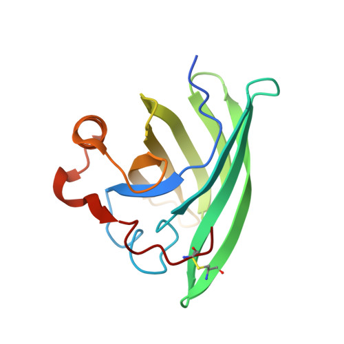
Crystal structure of a truncated form of porcine odorant-binding protein.

Perduca, M.Mancia, F.Del Giorgio, R.Monaco, H.L.

(2001) Proteins 42: 201-209

  • DOI: https://doi.org/10.1002/1097-0134(20010201)42:2<201::aid-prot70>3.0.co;2-7
  • Primary Citation of Related Structures:  
    1HQP

  • PubMed Abstract: 

    The odorant-binding proteins (OBPs) are a family of structurally related molecules that are found in high concentrations in the nasal mucus of vertebrates and bind with moderate affinity a large family of hydrophobic odorants. On the basis of their quaternary structure, the OBPs have been classified as monomers, homodimers, and heterodimers. Porcine OBP was believed for a long time to be a monomer under physiological conditions but there are recent data that support the existence of a monomer-dimer equilibrium. We have determined the crystal structure of a monoclinic form of porcine OBP and found that the truncated molecules, which lack the first 8 amino acids, pack in the cell as dimers that appear to have physiological relevance. The presence in the maps of electron density for an endogenous ligand has also let us identify the side chain of the amino acids that are at the ligand-binding site. In addition, an alternative way of access to the central cavity that binds the ligands is suggested by the particular packing of the molecules in this unit cell. Proteins 2001;42:201-209.


  • Organizational Affiliation

    Biocrystallography Laboratory, Department of Science and Technology, University of Verona, Italy.


Macromolecules
Find similar proteins by:  (by identity cutoff)  |  3D Structure
Entity ID: 1
MoleculeChains Sequence LengthOrganismDetailsImage
ODORANT-BINDING PROTEIN157Sus scrofaMutation(s): 0 
UniProt
Find proteins for P81245 (Sus scrofa)
Explore P81245 
Go to UniProtKB:  P81245
Entity Groups  
Sequence Clusters30% Identity50% Identity70% Identity90% Identity95% Identity100% Identity
UniProt GroupP81245
Sequence Annotations
Expand
  • Reference Sequence
Small Molecules
Ligands 1 Unique
IDChains Name / Formula / InChI Key2D Diagram3D Interactions
PRZ
Query on PRZ

Download Ideal Coordinates CCD File 
B [auth A]2-ISOBUTYL-3-METHOXYPYRAZINE
C9 H14 N2 O
UXFSPRAGHGMRSQ-UHFFFAOYSA-N
Experimental Data & Validation

Experimental Data

  • Method: X-RAY DIFFRACTION
  • Resolution: 2.30 Å
  • R-Value Work: 0.204 
  • R-Value Observed: 0.204 
  • Space Group: C 1 2 1
Unit Cell:
Length ( Å )Angle ( ˚ )
a = 89.18α = 90
b = 44.14β = 107.03
c = 41.24γ = 90
Software Package:
Software NamePurpose
X-GENdata scaling
X-GENdata reduction
AMoREphasing
TNTrefinement

Structure Validation

View Full Validation Report


Entry History 

Deposition Data

Revision History  (Full details and data files)

  • Version 1.0: 2001-01-17
    Type: Initial release
  • Version 1.1: 2008-04-27
    Changes: Version format compliance
  • Version 1.2: 2011-07-13
    Changes: Version format compliance
  • Version 1.3: 2017-10-04
    Changes: Refinement description
  • Version 1.4: 2023-08-09
    Changes: Data collection, Database references, Derived calculations, Refinement description