1H6O

Dimerisation domain from human TRF1


Experimental Data Snapshot

  • Method: X-RAY DIFFRACTION
  • Resolution: 2.90 Å
  • R-Value Free: 0.303 
  • R-Value Work: 0.250 
  • R-Value Observed: 0.250 

wwPDB Validation   3D Report Full Report


This is version 1.5 of the entry. See complete history


Literature

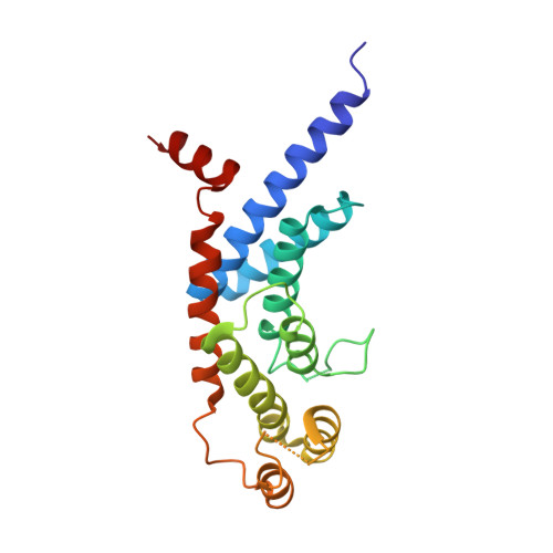
Structure of the TRFH dimerization domain of the human telomeric proteins TRF1 and TRF2.

Fairall, L.Chapman, L.Moss, H.de Lange, T.Rhodes, D.

(2001) Mol Cell 8: 351-361

  • DOI: https://doi.org/10.1016/s1097-2765(01)00321-5
  • Primary Citation of Related Structures:  
    1H6O, 1H6P

  • PubMed Abstract: 

    TRF1 and TRF2 are key components of vertebrate telomeres. They bind to double-stranded telomeric DNA as homodimers. Dimerization involves the TRF homology (TRFH) domain, which also mediates interactions with other telomeric proteins. The crystal structures of the dimerization domains from human TRF1 and TRF2 were determined at 2.9 and 2.2 A resolution, respectively. Despite a modest sequence identity, the two TRFH domains have the same entirely alpha-helical architecture, resembling a twisted horseshoe. The dimerization interfaces feature unique interactions that prevent heterodimerization. Mutational analysis of TRF1 corroborates the structural data and underscores the importance of the TRFH domain in dimerization, DNA binding, and telomere localization. A possible structural homology between the TRFH domain of fission yeast telomeric protein Taz1 with those of the vertebrate TRFs is suggested.


  • Organizational Affiliation

    MRC Laboratory of Molecular Biology, Hills Road, CB2 2QH, Cambridge, United Kingdom.


Macromolecules
Find similar proteins by:  (by identity cutoff)  |  3D Structure
Entity ID: 1
MoleculeChains Sequence LengthOrganismDetailsImage
TELOMERIC REPEAT BINDING FACTOR 1204Homo sapiensMutation(s): 0 
UniProt & NIH Common Fund Data Resources
Find proteins for P54274 (Homo sapiens)
Explore P54274 
Go to UniProtKB:  P54274
PHAROS:  P54274
GTEx:  ENSG00000147601 
Entity Groups  
Sequence Clusters30% Identity50% Identity70% Identity90% Identity95% Identity100% Identity
UniProt GroupP54274
Sequence Annotations
Expand
  • Reference Sequence
Experimental Data & Validation

Experimental Data

  • Method: X-RAY DIFFRACTION
  • Resolution: 2.90 Å
  • R-Value Free: 0.303 
  • R-Value Work: 0.250 
  • R-Value Observed: 0.250 
  • Space Group: P 31 2 1
Unit Cell:
Length ( Å )Angle ( ˚ )
a = 85.39α = 90
b = 85.39β = 90
c = 91.63γ = 120
Software Package:
Software NamePurpose
CNSrefinement
MOSFLMdata reduction
SCALAdata scaling
CCP4phasing
SOLVEphasing
SHARPphasing

Structure Validation

View Full Validation Report


Entry History 

Deposition Data

Revision History  (Full details and data files)

  • Version 1.0: 2001-09-05
    Type: Initial release
  • Version 1.1: 2011-05-08
    Changes: Version format compliance
  • Version 1.2: 2011-07-13
    Changes: Version format compliance
  • Version 1.3: 2018-06-13
    Changes: Data collection, Database references
  • Version 1.4: 2019-10-09
    Changes: Data collection, Database references, Other
  • Version 1.5: 2024-04-24
    Changes: Data collection, Database references