1FMB

EIAV PROTEASE COMPLEXED WITH THE INHIBITOR HBY-793


Experimental Data Snapshot

  • Method: X-RAY DIFFRACTION
  • Resolution: 1.80 Å
  • R-Value Work: 0.136 

wwPDB Validation   3D Report Full Report


This is version 1.5 of the entry. See complete history


Literature

Structure of equine infectious anemia virus proteinase complexed with an inhibitor.

Gustchina, A.Kervinen, J.Powell, D.J.Zdanov, A.Kay, J.Wlodawer, A.

(1996) Protein Sci 5: 1453-1465

  • DOI: https://doi.org/10.1002/pro.5560050802
  • Primary Citation of Related Structures:  
    1FMB

  • PubMed Abstract: 

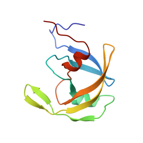
    Equine infectious anemia virus (EIAV), the causative agent of infectious anemia in horses, is a member of the lentiviral family. The virus-encoded proteinase (PR) processes viral polyproteins into functional molecules during replication and it also cleaves viral nucleocapsid protein during infection. The X-ray structure of a complex of the 154G mutant of EIAV PR with the inhibitor HBY-793 was solved at 1.8 A resolution and refined to a crystallographic R-factor of 0.136. The molecule is a dimer in which the monomers are related by a crystallographic twofold axis. Although both the enzyme and the inhibitor are symmetric, the interactions between the central part of the inhibitor and the active site aspartates are asymmetric, and the inhibitor and the two flaps are partially disordered. The overall fold of EIAV PR is very similar to that of other retroviral proteinases. However, a novel feature of the EIAV PR structure is the appearance of the second alpha-helix in the monomer in a position predicted by the structural template for the family of aspartic proteinases. The parts of the EIAV PR with the highest resemblance to human immunodeficiency virus type 1 PR include the substrate-binding sites; thus, the differences in the specificity of both enzymes have to be explained by enzyme-ligand interactions at the periphery of the active site as well.


  • Organizational Affiliation

    Macromolecular Structure Laboratory, NCI-Frederick Cancer Research and Development Center, Maryland 21702, USA.


Macromolecules
Find similar proteins by:  (by identity cutoff)  |  3D Structure
Entity ID: 1
MoleculeChains Sequence LengthOrganismDetailsImage
EIAV PROTEASE104Equine infectious anemia virusMutation(s): 1 
EC: 3.4.23.16
UniProt
Find proteins for P32542 (Equine infectious anemia virus (isolate CL22))
Explore P32542 
Go to UniProtKB:  P32542
Entity Groups  
Sequence Clusters30% Identity50% Identity70% Identity90% Identity95% Identity100% Identity
UniProt GroupP32542
Sequence Annotations
Expand
  • Reference Sequence
Small Molecules
Ligands 1 Unique
IDChains Name / Formula / InChI Key2D Diagram3D Interactions
HYB
Query on HYB

Download Ideal Coordinates CCD File 
B [auth A][2-(2-METHYL-PROPANE-2-SULFONYLMETHYL)-3-NAPHTHALEN-1-YL-PROPIONYL-VALINYL]-PHENYLALANINOL
C32 H42 N2 O5 S
DRAZKRRTKFXIQX-XQFUHLNNSA-N
Binding Affinity Annotations 
IDSourceBinding Affinity
HYB Binding MOAD:  1FMB Ki: 0.1 (nM) from 1 assay(s)
PDBBind:  1FMB Ki: 0.1 (nM) from 1 assay(s)
Experimental Data & Validation

Experimental Data

  • Method: X-RAY DIFFRACTION
  • Resolution: 1.80 Å
  • R-Value Work: 0.136 
  • Space Group: C 1 2 1
Unit Cell:
Length ( Å )Angle ( ˚ )
a = 42.93α = 90
b = 45.71β = 110.5
c = 56.77γ = 90
Software Package:
Software NamePurpose
X-PLORmodel building
X-PLORrefinement
DENZOdata reduction
SCALEPACKdata scaling
X-PLORphasing

Structure Validation

View Full Validation Report


Entry History 

Deposition Data

Revision History  (Full details and data files)

  • Version 1.0: 1996-10-14
    Type: Initial release
  • Version 1.1: 2008-03-24
    Changes: Version format compliance
  • Version 1.2: 2011-07-13
    Changes: Derived calculations, Version format compliance
  • Version 1.3: 2021-11-03
    Changes: Database references, Derived calculations, Other
  • Version 1.4: 2024-02-07
    Changes: Data collection
  • Version 1.5: 2024-04-03
    Changes: Refinement description