1FKX

MURINE ADENOSINE DEAMINASE (D296A)


Experimental Data Snapshot

  • Method: X-RAY DIFFRACTION
  • Resolution: 2.40 Å
  • R-Value Work: 0.158 
  • R-Value Observed: 0.158 

wwPDB Validation   3D Report Full Report


This is version 1.4 of the entry. See complete history


Literature

Probing the functional role of two conserved active site aspartates in mouse adenosine deaminase.

Sideraki, V.Mohamedali, K.A.Wilson, D.K.Chang, Z.Kellems, R.E.Quiocho, F.A.Rudolph, F.B.

(1996) Biochemistry 35: 7862-7872

  • DOI: https://doi.org/10.1021/bi952920d
  • Primary Citation of Related Structures:  
    1FKW, 1FKX

  • PubMed Abstract: 

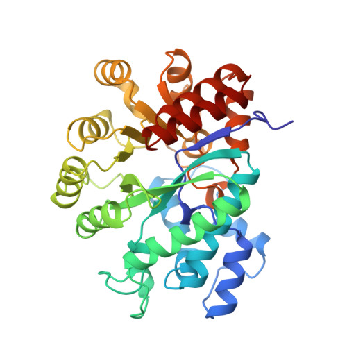
    Two adjacent aspartates, Asp 295 and Asp 296, playing major roles in the reaction catalyzed by mouse adenosine deaminase (mADA) were altered using site-directed mutagenesis. These mutants were expressed and purified from an ADA-deficient bacterial strain and characterized. Circular dichroism spectroscopy shows the mutants to have unperturbed secondary structure. Their zinc content compares well to that of wild-type enzyme. Changing Asp 295 to a glutamate decreases the kcat but does not alter the Km for adenosine, confirming the importance of this residue in the catalytic process and its minimal role in substrate binding. The crystal structure of the D295E mutant reveals a displacement of the catalytic water from the active site due to the longer glutamate side chain, resulting in the mutant's inability to turn over the substrate. In contrast, Asp 296 mutants exhibit markedly increased Km values, establishing this residue's critical role in substrate binding. The Asp 296->Ala mutation causes a 70-fold increase in the Km for adenosine and retains 0.001% of the wild-type kcat/Km value, whereas the ASP 296->Asn mutant has a 10-fold higher Km and retains 1% of the wild-type kcat/Km value. The structure of the D296A mutant shows that the impaired binding of substrate is caused by the loss of a single hydrogen bond between a carboxylate oxygen and N7 of the purine ring. These results and others discussed below are in agreement with the postulated role of the adjacent aspartates in the catalytic mechanism for mADA.


  • Organizational Affiliation

    Department of Biochemistry and Cell Biology, Rice University, Houston, Texas 77005, USA.


Macromolecules
Find similar proteins by:  (by identity cutoff)  |  3D Structure
Entity ID: 1
MoleculeChains Sequence LengthOrganismDetailsImage
ADENOSINE DEAMINASE349Mus musculusMutation(s): 1 
EC: 3.5.4.4
UniProt
Find proteins for P03958 (Mus musculus)
Explore P03958 
Go to UniProtKB:  P03958
Entity Groups  
Sequence Clusters30% Identity50% Identity70% Identity90% Identity95% Identity100% Identity
UniProt GroupP03958
Sequence Annotations
Expand
  • Reference Sequence
Small Molecules
Ligands 2 Unique
IDChains Name / Formula / InChI Key2D Diagram3D Interactions
PRH
Query on PRH

Download Ideal Coordinates CCD File 
C [auth A]6-HYDROXY-1,6-DIHYDRO PURINE NUCLEOSIDE
C10 H15 N4 O5
WGRXVKRHIMUTPD-YOHZANMFSA-O
ZN
Query on ZN

Download Ideal Coordinates CCD File 
B [auth A]ZINC ION
Zn
PTFCDOFLOPIGGS-UHFFFAOYSA-N
Binding Affinity Annotations 
IDSourceBinding Affinity
PRH Binding MOAD:  1FKX Ki: 6.00e+6 (nM) from 1 assay(s)
Experimental Data & Validation

Experimental Data

  • Method: X-RAY DIFFRACTION
  • Resolution: 2.40 Å
  • R-Value Work: 0.158 
  • R-Value Observed: 0.158 
  • Space Group: C 1 2 1
Unit Cell:
Length ( Å )Angle ( ˚ )
a = 102.71α = 90
b = 94.07β = 127.27
c = 73.08γ = 90
Software Package:
Software NamePurpose
X-PLORmodel building
X-PLORrefinement
SDMSdata reduction
X-PLORphasing

Structure Validation

View Full Validation Report


Entry History 

Deposition Data

Revision History  (Full details and data files)

  • Version 1.0: 1996-08-01
    Type: Initial release
  • Version 1.1: 2008-03-24
    Changes: Version format compliance
  • Version 1.2: 2011-07-13
    Changes: Version format compliance
  • Version 1.3: 2021-11-03
    Changes: Database references, Derived calculations, Other
  • Version 1.4: 2024-02-07
    Changes: Data collection