1F9F

CRYSTAL STRUCTURE OF THE HPV-18 E2 DNA-BINDING DOMAIN


Experimental Data Snapshot

  • Method: X-RAY DIFFRACTION
  • Resolution: 1.90 Å
  • R-Value Free: 0.294 
  • R-Value Work: 0.237 
  • R-Value Observed: 0.237 

wwPDB Validation   3D Report Full Report


This is version 1.4 of the entry. See complete history


Literature

The structural basis of DNA target discrimination by papillomavirus E2 proteins.

Kim, S.S.Tam, J.K.Wang, A.F.Hegde, R.S.

(2000) J Biol Chem 275: 31245-31254

  • DOI: https://doi.org/10.1074/jbc.M004541200
  • Primary Citation of Related Structures:  
    1F9F, 1JJ4

  • PubMed Abstract: 

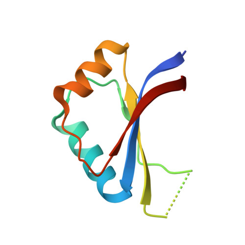
    The papillomavirus E2 proteins regulate the transcription of all papillomavirus genes and are necessary for viral DNA replication. Disruption of the E2 gene is commonly associated with malignancy in cervical carcinoma, indicating that E2 has a role in regulating tumor progression. Although the E2 proteins from all characterized papillomaviruses bind specifically to the same 12-base pair DNA sequence, the cancer-associated human papillomavirus E2 proteins display a unique ability to detect DNA flexibility and intrinsic curvature. To understand the structural basis for this phenomenon, we have determined the crystal structures of the human papillomavirus-18 E2 DNA-binding domain and its complexes with high and low affinity binding sites. The E2 protein is a dimeric beta-barrel and the E2-DNA interaction is accompanied by a large deformation of the DNA as it conforms to the E2 surface. DNA conformation and E2-DNA contacts are similar in both high and low affinity complexes. The differences in affinity correlate with the flexibility of the DNA sequence. Preferences of E2 proteins from different papillomavirus strains for flexible or prevent DNA targets correlate with the distribution of positive charge on their DNA interaction surfaces, suggesting a role for electrostatic forces in the recognition of DNA deformability.


  • Organizational Affiliation

    Department of Biochemistry and Program in Structural Biology, New York University Medical Center, Skirball Institute of Biomolecular Medicine, New York, New York 10016, USA.


Macromolecules
Find similar proteins by:  (by identity cutoff)  |  3D Structure
Entity ID: 1
MoleculeChains Sequence LengthOrganismDetailsImage
Regulatory protein E2
A, B, C, D
83human papillomavirus 18Mutation(s): 0 
Gene Names: E2
UniProt
Find proteins for P06790 (Human papillomavirus type 18)
Explore P06790 
Go to UniProtKB:  P06790
Entity Groups  
Sequence Clusters30% Identity50% Identity70% Identity90% Identity95% Identity100% Identity
UniProt GroupP06790
Sequence Annotations
Expand
  • Reference Sequence
Small Molecules
Ligands 1 Unique
IDChains Name / Formula / InChI Key2D Diagram3D Interactions
SO4
Query on SO4

Download Ideal Coordinates CCD File 
E [auth C],
F [auth D]
SULFATE ION
O4 S
QAOWNCQODCNURD-UHFFFAOYSA-L
Experimental Data & Validation

Experimental Data

  • Method: X-RAY DIFFRACTION
  • Resolution: 1.90 Å
  • R-Value Free: 0.294 
  • R-Value Work: 0.237 
  • R-Value Observed: 0.237 
  • Space Group: P 21 21 21
Unit Cell:
Length ( Å )Angle ( ˚ )
a = 41.5α = 90
b = 46.8β = 90
c = 161.06γ = 90
Software Package:
Software NamePurpose
AMoREphasing
CNSrefinement
DENZOdata reduction
SCALEPACKdata scaling

Structure Validation

View Full Validation Report


Entry History 

Deposition Data

Revision History  (Full details and data files)

  • Version 1.0: 2000-11-15
    Type: Initial release
  • Version 1.1: 2008-04-27
    Changes: Version format compliance
  • Version 1.2: 2011-07-13
    Changes: Version format compliance
  • Version 1.3: 2018-06-13
    Changes: Advisory, Data collection, Database references, Source and taxonomy, Structure summary
  • Version 1.4: 2024-02-07
    Changes: Data collection, Database references, Derived calculations