1F76

ESCHERICHIA COLI DIHYDROOROTATE DEHYDROGENASE


Experimental Data Snapshot

  • Method: X-RAY DIFFRACTION
  • Resolution: 2.50 Å
  • R-Value Free: 0.217 
  • R-Value Work: 0.177 

wwPDB Validation   3D Report Full Report


Ligand Structure Quality Assessment 


This is version 1.3 of the entry. See complete history


Literature

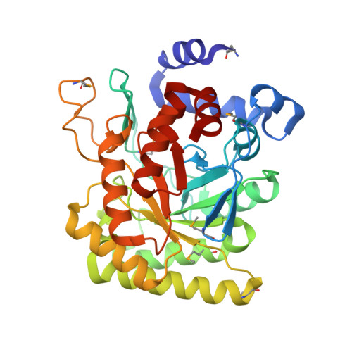
E. coli Dihydroorotate Dehydrogenase Reveals Structural and Functional Distinction between different classes of dihydroorotate dehydrogenases

Norager, S.Jensen, K.F.Bjornberg, O.Larsen, S.

(2002) Structure 10: 1211-1223

  • DOI: https://doi.org/10.1016/s0969-2126(02)00831-6
  • Primary Citation of Related Structures:  
    1F76

  • PubMed Abstract: 

    The flavoenzymes dihydroorotate dehydrogenases (DHODs) catalyze the fourth and only redox step in the de novo biosynthesis of UMP. Enzymes belonging to class 2, according to their amino acid sequence, are characterized by having a serine residue as the catalytic base and a longer N terminus. The structure of class 2 E. coli DHOD, determined by MAD phasing, showed that the N-terminal extension forms a separate domain. The catalytic serine residue has an environment differing from the equivalent cysteine in class 1 DHODs. Significant differences between the two classes of DHODs were identified by comparison of the E. coli DHOD with the other known DHOD structures, and differences with the class 2 human DHOD explain the variation in their inhibitors.


  • Organizational Affiliation

    Centre for Crystallographic Studies, University of Copenhagen, Universitetsparken 5, DK-2100 Copenhagen, Denmark.


Macromolecules
Find similar proteins by:  (by identity cutoff)  |  3D Structure
Entity ID: 1
MoleculeChains Sequence LengthOrganismDetailsImage
Dihydroorotate dehydrogenase (quinone)A,
B,
C [auth D],
D [auth E]
336Escherichia coli K-12Mutation(s): 0 
Gene Names: pyrDb0945JW0928
EC: 1.3.5.2
Membrane Entity: Yes 
UniProt
Find proteins for P0A7E1 (Escherichia coli (strain K12))
Explore P0A7E1 
Go to UniProtKB:  P0A7E1
Entity Groups  
Sequence Clusters30% Identity50% Identity70% Identity90% Identity95% Identity100% Identity
UniProt GroupP0A7E1
Sequence Annotations
Expand
  • Reference Sequence
Small Molecules
Ligands 3 Unique
IDChains Name / Formula / InChI Key2D Diagram3D Interactions
FMN
Query on FMN

Download Ideal Coordinates CCD File 
E [auth A],
H [auth B],
L [auth D],
P [auth E]
FLAVIN MONONUCLEOTIDE
C17 H21 N4 O9 P
FVTCRASFADXXNN-SCRDCRAPSA-N
ORO
Query on ORO

Download Ideal Coordinates CCD File 
F [auth A],
G [auth A],
M [auth D]
OROTIC ACID
C5 H4 N2 O4
PXQPEWDEAKTCGB-UHFFFAOYSA-N
FMT
Query on FMT

Download Ideal Coordinates CCD File 
I [auth B]
J [auth B]
K [auth B]
N [auth D]
O [auth D]
I [auth B],
J [auth B],
K [auth B],
N [auth D],
O [auth D],
Q [auth E],
R [auth E],
S [auth E]
FORMIC ACID
C H2 O2
BDAGIHXWWSANSR-UHFFFAOYSA-N
Modified Residues  1 Unique
IDChains TypeFormula2D DiagramParent
MSE
Query on MSE
A,
B,
C [auth D],
D [auth E]
L-PEPTIDE LINKINGC5 H11 N O2 SeMET
Experimental Data & Validation

Experimental Data

  • Method: X-RAY DIFFRACTION
  • Resolution: 2.50 Å
  • R-Value Free: 0.217 
  • R-Value Work: 0.177 
  • Space Group: P 41 21 2
Unit Cell:
Length ( Å )Angle ( ˚ )
a = 119.7α = 90
b = 119.7β = 90
c = 295.97γ = 90
Software Package:
Software NamePurpose
DENZOdata reduction
SCALEPACKdata scaling
SOLVEphasing
CNSrefinement

Structure Validation

View Full Validation Report


Ligand Structure Quality Assessment 


Entry History 

Deposition Data

Revision History  (Full details and data files)

  • Version 1.0: 2002-10-16
    Type: Initial release
  • Version 1.1: 2008-04-27
    Changes: Version format compliance
  • Version 1.2: 2011-07-13
    Changes: Version format compliance
  • Version 1.3: 2014-03-12
    Changes: Source and taxonomy, Version format compliance