Type II protein secretion in gram-negative pathogenic bacteria: the study of the structure/secretion relationships of the cellulase Cel5 (formerly EGZ) from Erwinia chrysanthemi
Chapon, M., Czjzek, M., El Hassouni, M., Py, B., Juy, M., Barras, F.(2001) J Mol Biol 310: 1055-1066
- PubMed: 11501995
- DOI: https://doi.org/10.1006/jmbi.2001.4787
- Primary Citation of Related Structures:
1EGZ - PubMed Abstract:
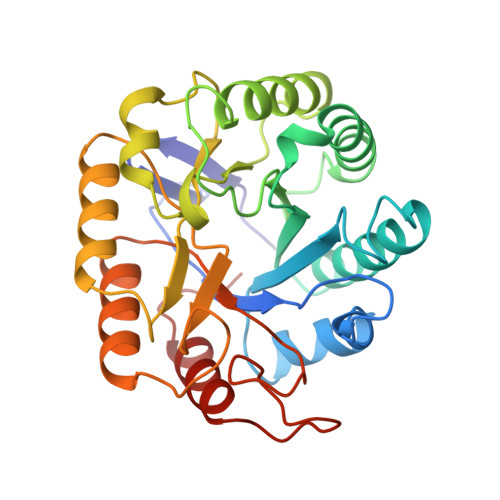
Erwinia chrysanthemi, a Gram-negative plant pathogen, secretes the cellulase Cel5 (formerly EGZ) via the type II secretion pathway (referred to as Out). Cel5 is composed of two domains, a large N-terminal catalytic domain (390 amino acid residues) and a small C-terminal cellulose-binding domain (62 amino acid residues) separated by a linker region. A combination of mutagenesis and structural analysis permitted us to investigate the structure/secretion relationships with respect to the catalytic domain of Cel5. The 3D structure of the catalytic domain was solved by molecular replacement at 2.3 A resolution. Cel5 exhibits the (beta/alpha)8 structural fold and two extra-barrel features. Our previous genetic study based upon tRNA-mediated suppression allowed us to predict positions of importance in the molecule in relation to structure and catalysis. Remarkably, all of the predictions proved to be correct when compared with the present structural information. Mutations of Arg57, which is located at the heart of the catalytic domain, allowed us to test the consequences of structural modifications on the secretion efficiency. The results revealed that secretability imposes remarkably strong constraints upon folding. In particular, an Arg-to-His mutation yielded a species that folded to a stable conformation close to, but distinct from the wild-type, which however was not secretable. We discuss the relationships between folding of a protein in the periplasm, en route to the cell exterior, and presentation of secretion information. We propose that different solutions have been selected for type II secreted exoproteins in order to meet the constraints imposed by their interaction with their respective secretion machineries. We propose that evolutionary pressure has led to the adaptation of different secretion motifs for different type II exoproteins.
Organizational Affiliation:
Laboratoire de Chimie Bactérienne , Institut de Biologie Structurale et Microbiologie CNRS-Marseille, France.