1ED8

STRUCTURE OF E. COLI ALKALINE PHOSPHATASE INHIBITED BY THE INORGANIC PHOSPHATE AT 1.75A RESOLUTION


Experimental Data Snapshot

  • Method: X-RAY DIFFRACTION
  • Resolution: 1.75 Å
  • R-Value Free: 0.228 
  • R-Value Work: 0.196 
  • R-Value Observed: 0.197 

wwPDB Validation   3D Report Full Report


This is version 1.2 of the entry. See complete history


Literature

A revised mechanism for the alkaline phosphatase reaction involving three metal ions.

Stec, B.Holtz, K.M.Kantrowitz, E.R.

(2000) J Mol Biol 299: 1303-1311

  • DOI: https://doi.org/10.1006/jmbi.2000.3799
  • Primary Citation of Related Structures:  
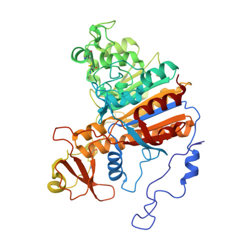
    1ED8, 1ED9

  • PubMed Abstract: 

    Here, X-ray crystallography has been used to investigate the proposed double in-line displacement mechanism of Escherichia coli alkaline phosphatase in which two of the three active-site metal ions have a direct role in catalysis. Two new X-ray crystal structures of the wild-type enzyme in the absence and presence of inorganic phosphate have been refined at 1.75 A to final working R-factors of 15.4% and 16.4%, respectively. In the refinement of both structures, residues in the active sites were treated anisotropically. The ellipsoids resulting from the partial anisotropic refinement show a clear route for the binding and release of substrate/product. In addition, a direct comparison of the refined structures with and without phosphate reveal a strong correlation between the occupancy of the third metal-binding site and the conformation of the Ser102 nucleophile. These findings clarify two important and unresolved aspects of the previously proposed catalytic mechanism, how Ser102 is activated for nucleophilic attack and why a magnesium ion in the third metal site is required for catalysis. Analysis of these results suggest that three metal-ion assisted catalysis is a more accurate description of the mechanism of the alkaline phosphatase reaction. A revised mechanism for the catalytic reaction of alkaline phosphatase is proposed on the basis of the two new X-ray crystal structures reported.


  • Organizational Affiliation

    Department of Biochemistry and Cell Biology, W. M. Keck Center for Computational Biology, Rice University, Houston, TX, 77005, USA.


Macromolecules
Find similar proteins by:  (by identity cutoff)  |  3D Structure
Entity ID: 1
MoleculeChains Sequence LengthOrganismDetailsImage
ALKALINE PHOSPHATASE
A, B
449Escherichia coliMutation(s): 0 
EC: 3.1.3.1
UniProt
Find proteins for P00634 (Escherichia coli (strain K12))
Explore P00634 
Go to UniProtKB:  P00634
Entity Groups  
Sequence Clusters30% Identity50% Identity70% Identity90% Identity95% Identity100% Identity
UniProt GroupP00634
Sequence Annotations
Expand
  • Reference Sequence
Small Molecules
Ligands 4 Unique
IDChains Name / Formula / InChI Key2D Diagram3D Interactions
SO4
Query on SO4

Download Ideal Coordinates CCD File 
H [auth A],
N [auth B]
SULFATE ION
O4 S
QAOWNCQODCNURD-UHFFFAOYSA-L
PO4
Query on PO4

Download Ideal Coordinates CCD File 
G [auth A],
M [auth B]
PHOSPHATE ION
O4 P
NBIIXXVUZAFLBC-UHFFFAOYSA-K
ZN
Query on ZN

Download Ideal Coordinates CCD File 
C [auth A]
D [auth A]
E [auth A]
I [auth B]
J [auth B]
C [auth A],
D [auth A],
E [auth A],
I [auth B],
J [auth B],
K [auth B]
ZINC ION
Zn
PTFCDOFLOPIGGS-UHFFFAOYSA-N
MG
Query on MG

Download Ideal Coordinates CCD File 
F [auth A],
L [auth B]
MAGNESIUM ION
Mg
JLVVSXFLKOJNIY-UHFFFAOYSA-N
Experimental Data & Validation

Experimental Data

  • Method: X-RAY DIFFRACTION
  • Resolution: 1.75 Å
  • R-Value Free: 0.228 
  • R-Value Work: 0.196 
  • R-Value Observed: 0.197 
  • Space Group: I 2 2 2
Unit Cell:
Length ( Å )Angle ( ˚ )
a = 195.15α = 90
b = 167.34β = 90
c = 76.8γ = 90
Software Package:
Software NamePurpose
SDMSdata collection
SDMSdata reduction
X-PLORmodel building
SHELXL-97refinement
SDMSdata scaling
X-PLORphasing

Structure Validation

View Full Validation Report


Entry History 

Deposition Data

Revision History  (Full details and data files)

  • Version 1.0: 2000-09-20
    Type: Initial release
  • Version 1.1: 2008-04-27
    Changes: Version format compliance
  • Version 1.2: 2011-07-13
    Changes: Version format compliance