1E2R

CYTOCHROME CD1 NITRITE REDUCTASE, REDUCED AND CYANIDE BOUND


Experimental Data Snapshot

  • Method: X-RAY DIFFRACTION
  • Resolution: 1.59 Å
  • R-Value Free: 0.208 
  • R-Value Work: 0.192 
  • R-Value Observed: 0.192 

wwPDB Validation   3D Report Full Report


This is version 1.3 of the entry. See complete history


Literature

X-Ray Crystallographic Study of Cyanide Binding Provides Insights Into the Structure-Function Relationship for Cytochrome Cd1 Nitrite Reductase from Paracoccus Pantotrophus.

Jafferji, A.Allen, J.W.Ferguson, S.J.Fulop, V.

(2000) J Biol Chem 275: 25089

  • DOI: https://doi.org/10.1074/jbc.M001377200
  • Primary Citation of Related Structures:  
    1E2R

  • PubMed Abstract: 

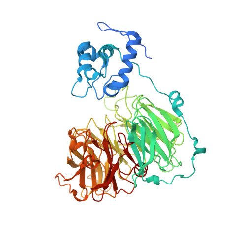
    We present a 1.59-A resolution crystal structure of reduced Paracoccus pantotrophus cytochrome cd(1) with cyanide bound to the d(1) heme and His/Met coordination of the c heme. Fe-C-N bond angles are 146 degrees for the A subunit and 164 degrees for the B subunit of the dimer. The nitrogen atom of bound cyanide is within hydrogen bonding distance of His(345) and His(388) and either a water molecule in subunit A or Tyr(25) in subunit B. The ferrous heme-cyanide complex is unusually stable (K(d) approximately 10(-6) m); we propose that this reflects both the design of the specialized d(1) heme ring and a general feature of anion reductases with active site heme. Oxidation of crystals of reduced, cyanide-bound, cytochrome cd(1) results in loss of cyanide and return to the native structure with Tyr(25) as a ligand to the d(1) heme iron and switching to His/His coordination at the c-type heme. No reason for unusually weak binding of cyanide to the ferric state can be identified; rather it is argued that the protein is designed such that a chelate-based effect drives displacement by tyrosine of cyanide or a weaker ligand, like reaction product nitric oxide, from the ferric d(1) heme.


  • Organizational Affiliation

    Laboratory of Molecular Biophysics, Department of Biochemistry, University of Oxford, United Kingdom.


Macromolecules
Find similar proteins by:  (by identity cutoff)  |  3D Structure
Entity ID: 1
MoleculeChains Sequence LengthOrganismDetailsImage
NITRITE REDUCTASE
A, B
567Paracoccus denitrificansMutation(s): 0 
EC: 1.7.2.1 (PDB Primary Data), 1.7.99.1 (PDB Primary Data)
UniProt
Find proteins for P72181 (Paracoccus pantotrophus)
Explore P72181 
Go to UniProtKB:  P72181
Entity Groups  
Sequence Clusters30% Identity50% Identity70% Identity90% Identity95% Identity100% Identity
UniProt GroupP72181
Sequence Annotations
Expand
  • Reference Sequence
Experimental Data & Validation

Experimental Data

  • Method: X-RAY DIFFRACTION
  • Resolution: 1.59 Å
  • R-Value Free: 0.208 
  • R-Value Work: 0.192 
  • R-Value Observed: 0.192 
  • Space Group: P 1 21 1
Unit Cell:
Length ( Å )Angle ( ˚ )
a = 106.5α = 90
b = 60.9β = 111.9
c = 100.1γ = 90
Software Package:
Software NamePurpose
X-PLORrefinement
DENZOdata reduction
SCALEPACKdata scaling
CCP4phasing
X-PLORphasing
Ophasing

Structure Validation

View Full Validation Report


Entry History 

Deposition Data

  • Released Date: 2000-05-30 
  • Deposition Author(s): Fulop, V.

Revision History  (Full details and data files)

  • Version 1.0: 2000-05-30
    Type: Initial release
  • Version 1.1: 2011-10-19
    Changes: Data collection, Derived calculations, Non-polymer description, Other, Structure summary, Version format compliance
  • Version 1.2: 2019-07-24
    Changes: Data collection, Derived calculations
  • Version 1.3: 2023-12-06
    Changes: Data collection, Database references, Derived calculations, Refinement description