1DVO

THE X-RAY CRYSTAL STRUCTURE OF FINO, A REPRESSOR OF BACTERIAL CONJUGATION


Experimental Data Snapshot

  • Method: X-RAY DIFFRACTION
  • Resolution: 2.00 Å
  • R-Value Free: 0.224 
  • R-Value Work: 0.197 

wwPDB Validation   3D Report Full Report


This is version 1.4 of the entry. See complete history


Literature

Crystal structure of the bacterial conjugation repressor finO.

Ghetu, A.F.Gubbins, M.J.Frost, L.S.Glover, J.N.

(2000) Nat Struct Biol 7: 565-569

  • DOI: https://doi.org/10.1038/76790
  • Primary Citation of Related Structures:  
    1DVO

  • PubMed Abstract: 

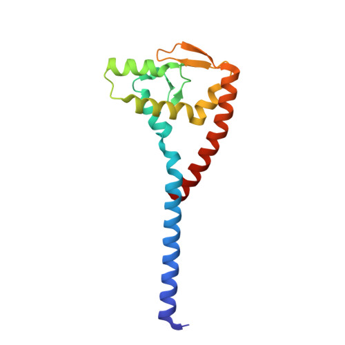
    The conjugative transfer of F-like plasmids is repressed by FinO, an RNA binding protein. FinO interacts with the F-plasmid encoded traJ mRNA and its antisense RNA, FinP, stabilizing FinP against endonucleolytic degradation and facilitating sense-antisense RNA recognition. Here we present the 2.0 A resolution X-ray crystal structure of FinO, lacking its flexible N-terminal extension. FinO adopts a novel, elongated, largely helical conformation. An N-terminal region, previously shown to contact RNA, forms a positively charged alpha-helix (helix 1) that protrudes 45 A from the central core of FinO. A C-terminal region of FinO that is implicated in RNA interactions also extends out from the central body of the protein, adopting a helical conformation and packing against the base of the N-terminal helix. A highly positively charged patch on the surface of the FinO core may present another RNA binding surface. The results of an in vitro RNA duplexing assay demonstrate that the flexible N-terminal region of FinO plays a key role in FinP-traJ RNA recognition, and supports our proposal that this region and the N-terminus of helix 1 interact with and stabilize paired, complementary RNA loops in a kissing complex.


  • Organizational Affiliation

    Department of Biochemistry, University of Alberta, Edmonton, Alberta, T6G 2H7, Canada.


Macromolecules
Find similar proteins by:  (by identity cutoff)  |  3D Structure
Entity ID: 1
MoleculeChains Sequence LengthOrganismDetailsImage
FERTILITY INHIBITION PROTEIN O152Escherichia coliMutation(s): 1 
UniProt
Find proteins for P29367 (Escherichia coli)
Explore P29367 
Go to UniProtKB:  P29367
Entity Groups  
Sequence Clusters30% Identity50% Identity70% Identity90% Identity95% Identity100% Identity
UniProt GroupP29367
Sequence Annotations
Expand
  • Reference Sequence
Experimental Data & Validation

Experimental Data

  • Method: X-RAY DIFFRACTION
  • Resolution: 2.00 Å
  • R-Value Free: 0.224 
  • R-Value Work: 0.197 
  • Space Group: P 21 21 21
Unit Cell:
Length ( Å )Angle ( ˚ )
a = 37.57α = 90
b = 38.726β = 90
c = 145.42γ = 90
Software Package:
Software NamePurpose
SOLVEphasing
CNSrefinement
DENZOdata reduction
SCALEPACKdata scaling

Structure Validation

View Full Validation Report


Entry History 

Deposition Data

Revision History  (Full details and data files)

  • Version 1.0: 2000-07-19
    Type: Initial release
  • Version 1.1: 2008-04-27
    Changes: Version format compliance
  • Version 1.2: 2011-07-13
    Changes: Version format compliance
  • Version 1.3: 2021-11-03
    Changes: Database references
  • Version 1.4: 2024-02-07
    Changes: Data collection