1DK7

CRYSTAL STRUCTURE OF AN ISOLATED APICAL DOMAIN OF GROEL


Experimental Data Snapshot

  • Method: X-RAY DIFFRACTION
  • Resolution: 2.02 Å
  • R-Value Free: 0.241 
  • R-Value Work: 0.225 
  • R-Value Observed: 0.225 

wwPDB Validation   3D Report Full Report


This is version 1.4 of the entry. See complete history


Literature

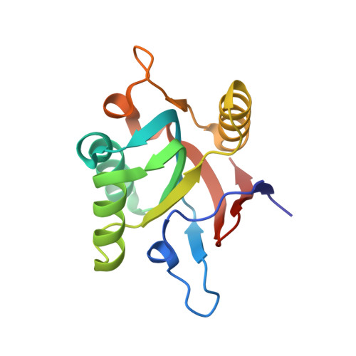
The crystal structure of a GroEL/peptide complex: plasticity as a basis for substrate diversity.

Chen, L.Sigler, P.B.

(1999) Cell 99: 757-768

  • DOI: https://doi.org/10.1016/s0092-8674(00)81673-6
  • Primary Citation of Related Structures:  
    1DK7, 1DKD

  • PubMed Abstract: 

    The chaperonin GroEL is a double toriodal assembly that with its cochaperonin GroES facilitates protein folding with an ATP-dependent mechanism. Nonnative conformations of diverse protein substrates bind to the apical domains surrounding the opening of the double toroid's central cavity. Using phage display, we have selected peptides with high affinity for the isolated apical domain. We have determined the crystal structures of the complexes formed by the most strongly bound peptide with the isolated apical domain, and with GroEL. The peptide interacts with the groove between paired alpha helices in a manner similar to that of the GroES mobile loop. Our structural analysis, combined with other results, suggests that various modes of molecular plasticity are responsible for tight promiscuous binding of nonnative substrates and their release into the shielded cis assembly.


  • Organizational Affiliation

    Department of Molecular Biophysics and Biochemistry, Yale University, New Haven, Connecticut 06511, USA.


Macromolecules
Find similar proteins by:  (by identity cutoff)  |  3D Structure
Entity ID: 1
MoleculeChains Sequence LengthOrganismDetailsImage
GROEL
A, B
146Escherichia coliMutation(s): 0 
UniProt
Find proteins for P0A6F5 (Escherichia coli (strain K12))
Explore P0A6F5 
Go to UniProtKB:  P0A6F5
Entity Groups  
Sequence Clusters30% Identity50% Identity70% Identity90% Identity95% Identity100% Identity
UniProt GroupP0A6F5
Sequence Annotations
Expand
  • Reference Sequence
Experimental Data & Validation

Experimental Data

  • Method: X-RAY DIFFRACTION
  • Resolution: 2.02 Å
  • R-Value Free: 0.241 
  • R-Value Work: 0.225 
  • R-Value Observed: 0.225 
  • Space Group: P 32 2 1
Unit Cell:
Length ( Å )Angle ( ˚ )
a = 84.859α = 90
b = 84.859β = 90
c = 77.068γ = 120
Software Package:
Software NamePurpose
CNSrefinement
DENZOdata reduction
SCALEPACKdata scaling
CNSphasing

Structure Validation

View Full Validation Report


Entry History 

Deposition Data

Revision History  (Full details and data files)

  • Version 1.0: 2000-01-05
    Type: Initial release
  • Version 1.1: 2008-04-27
    Changes: Version format compliance
  • Version 1.2: 2011-07-13
    Changes: Version format compliance
  • Version 1.3: 2018-01-31
    Changes: Experimental preparation
  • Version 1.4: 2024-02-07
    Changes: Data collection, Database references