1D5N

CRYSTAL STRUCTURE OF E. COLI MNSOD AT 100K


Experimental Data Snapshot

  • Method: X-RAY DIFFRACTION
  • Resolution: 1.55 Å
  • R-Value Free: 0.233 
  • R-Value Work: 0.194 
  • R-Value Observed: 0.194 

wwPDB Validation   3D Report Full Report


This is version 1.4 of the entry. See complete history


Literature

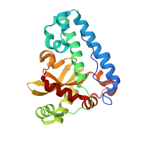
Cryo-trapping the six-coordinate, distorted-octahedral active site of manganese superoxide dismutase.

Borgstahl, G.E.Pokross, M.Chehab, R.Sekher, A.Snell, E.H.

(2000) J Mol Biol 296: 951-959

  • DOI: https://doi.org/10.1006/jmbi.1999.3506
  • Primary Citation of Related Structures:  
    1D5N

  • PubMed Abstract: 

    Superoxide dismutase protects organisms from potentially damaging oxygen radicals by catalyzing the disproportionation of superoxide to oxygen and hydrogen peroxide. We report the use of cryogenic temperatures to kinetically capture the sixth ligand bound to the active site of manganese superoxide dismutase (MnSOD). Synchrotron X-ray diffraction data was collected from Escherichia coli MnSOD crystals grown at pH 8.5 and cryocooled to 100 K. Structural refinement to 1.55 A resolution and close inspection of the active site revealed electron density for a sixth ligand that was interpreted to be a hydroxide ligand. The six-coordinate, distorted-octahedral geometry assumed during inhibition by hydroxide is compared to the room temperature, five-coordinate, trigonal bipyramidal active site determined with crystals grown from practically identical conditions. The gateway residues Tyr34, His30 and a tightly bound water molecule are implicated in closing-off the active site and blocking the escape route of the sixth ligand.


  • Organizational Affiliation

    Department of Chemistry, The University of Toledo, 2801 West Bancroft Street, Toledo, OH 43606, USA. gborgst@uoft02.utoledo.edu


Macromolecules
Find similar proteins by:  (by identity cutoff)  |  3D Structure
Entity ID: 1
MoleculeChains Sequence LengthOrganismDetailsImage
PROTEIN (MANGANESE SUPEROXIDE DISMUTASE)
A, B, C, D
205Escherichia coliMutation(s): 0 
EC: 1.15.1.1
UniProt
Find proteins for P00448 (Escherichia coli (strain K12))
Explore P00448 
Go to UniProtKB:  P00448
Entity Groups  
Sequence Clusters30% Identity50% Identity70% Identity90% Identity95% Identity100% Identity
UniProt GroupP00448
Sequence Annotations
Expand
  • Reference Sequence
Experimental Data & Validation

Experimental Data

  • Method: X-RAY DIFFRACTION
  • Resolution: 1.55 Å
  • R-Value Free: 0.233 
  • R-Value Work: 0.194 
  • R-Value Observed: 0.194 
  • Space Group: C 2 2 21
Unit Cell:
Length ( Å )Angle ( ˚ )
a = 99.11α = 90
b = 107.3β = 90
c = 179.11γ = 90
Software Package:
Software NamePurpose
X-PLORmodel building
X-PLORrefinement
MOSFLMdata reduction
CCP4data scaling
X-PLORphasing

Structure Validation

View Full Validation Report


Entry History 

Deposition Data

Revision History  (Full details and data files)

  • Version 1.0: 2000-03-02
    Type: Initial release
  • Version 1.1: 2008-04-27
    Changes: Version format compliance
  • Version 1.2: 2011-07-13
    Changes: Version format compliance
  • Version 1.3: 2011-11-16
    Changes: Atomic model
  • Version 1.4: 2024-02-07
    Changes: Data collection, Database references, Derived calculations