1CZQ

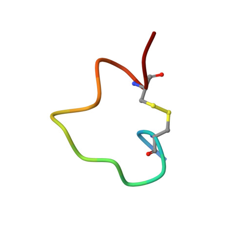
CRYSTAL STRUCTURE OF THE D10-P1/IQN17 COMPLEX: A D-PEPTIDE INHIBITOR OF HIV-1 ENTRY BOUND TO THE GP41 COILED-COIL POCKET.


Experimental Data Snapshot

  • Method: X-RAY DIFFRACTION
  • Resolution: 1.50 Å
  • R-Value Free: 0.245 
  • R-Value Work: 0.214 
  • R-Value Observed: 0.214 

wwPDB Validation   3D Report Full Report


This is version 1.2 of the entry. See complete history


Literature

Inhibiting HIV-1 entry: discovery of D-peptide inhibitors that target the gp41 coiled-coil pocket.

Eckert, D.M.Malashkevich, V.N.Hong, L.H.Carr, P.A.Kim, P.S.

(1999) Cell 99: 103-115

  • DOI: https://doi.org/10.1016/s0092-8674(00)80066-5
  • Primary Citation of Related Structures:  
    1CZQ, 2Q3I, 2Q5U, 2Q7C

  • PubMed Abstract: 

    The HIV-1 gp41 protein promotes viral entry by mediating the fusion of viral and cellular membranes. A prominent pocket on the surface of a central trimeric coiled coil within gp41 was previously identified as a potential target for drugs that inhibit HIV-1 entry. We designed a peptide, IQN17, which properly presents this pocket. Utilizing IQN17 and mirror-image phage display, we identified cyclic, D-peptide inhibitors of HIV-1 infection that share a sequence motif. A 1.5 A cocrystal structure of IQN17 in complex with a D-peptide, and NMR studies, show that conserved residues of these inhibitors make intimate contact with the gp41 pocket. Our studies validate the pocket per se as a target for drug development. IQN17 and these D-peptide inhibitors are likely to be useful for development and identification of a new class of orally bioavailable anti-HIV drugs.


  • Organizational Affiliation

    Howard Hughes Medical Institute, Whitehead Institute for Biomedical Research, Department of Biology, Massachusetts Institute of Technology, Cambridge 02142, USA.


Macromolecules
Find similar proteins by:  (by identity cutoff)  |  3D Structure
Entity ID: 1
MoleculeChains Sequence LengthOrganismDetailsImage
FUSION PROTEIN BETWEEN THE HYDROPHOBIC POCKET OF HIV GP41 AND GCN4-PIQI46Saccharomyces cerevisiaeHuman immunodeficiency virus
This entity is chimeric
Mutation(s): 0 
Entity Groups  
Sequence Clusters30% Identity50% Identity70% Identity90% Identity95% Identity100% Identity
Sequence Annotations
Expand
  • Reference Sequence

Find similar proteins by:  Sequence   |   3D Structure  

Entity ID: 2
MoleculeChains Sequence LengthOrganismDetailsImage
D-PEPTIDE INHIBITORB [auth D]17N/AMutation(s): 0 
Entity Groups  
Sequence Clusters30% Identity50% Identity70% Identity90% Identity95% Identity100% Identity
Sequence Annotations
Expand
  • Reference Sequence
Small Molecules
Ligands 1 Unique
IDChains Name / Formula / InChI Key2D Diagram3D Interactions
CL
Query on CL

Download Ideal Coordinates CCD File 
C [auth A]CHLORIDE ION
Cl
VEXZGXHMUGYJMC-UHFFFAOYSA-M
Experimental Data & Validation

Experimental Data

  • Method: X-RAY DIFFRACTION
  • Resolution: 1.50 Å
  • R-Value Free: 0.245 
  • R-Value Work: 0.214 
  • R-Value Observed: 0.214 
  • Space Group: P 3 2 1
Unit Cell:
Length ( Å )Angle ( ˚ )
a = 41.829α = 90
b = 41.829β = 90
c = 84.817γ = 120
Software Package:
Software NamePurpose
DENZOdata reduction
SCALEPACKdata scaling
MLPHAREphasing
CNSrefinement

Structure Validation

View Full Validation Report


Entry History 

Deposition Data

Revision History  (Full details and data files)

  • Version 1.0: 1999-10-13
    Type: Initial release
  • Version 1.1: 2008-04-27
    Changes: Version format compliance
  • Version 1.2: 2011-07-13
    Changes: Atomic model, Database references, Derived calculations, Non-polymer description, Structure summary, Version format compliance