1CGT

STRUCTURE OF CYCLODEXTRIN GLYCOSYLTRANSFERASE REFINED AT 2.0 ANGSTROMS RESOLUTION


Experimental Data Snapshot

  • Method: X-RAY DIFFRACTION
  • Resolution: 2.00 Å
  • R-Value Work: 0.166 
  • R-Value Observed: 0.166 

wwPDB Validation   3D Report Full Report


This is version 1.2 of the entry. See complete history


Literature

Structure of cyclodextrin glycosyltransferase refined at 2.0 A resolution.

Klein, C.Schulz, G.E.

(1991) J Mol Biol 217: 737-750

  • DOI: https://doi.org/10.1016/0022-2836(91)90530-j
  • Primary Citation of Related Structures:  
    1CGT

  • PubMed Abstract: 

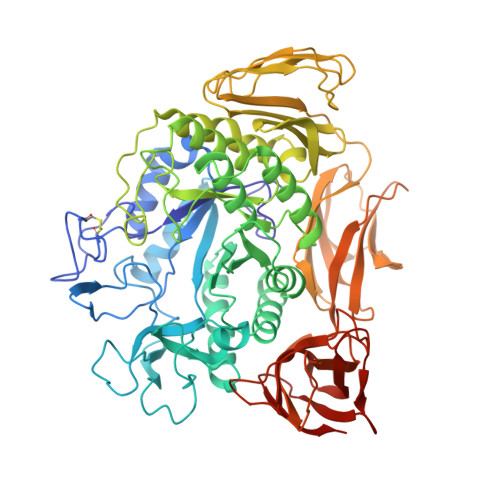
    The previously reported structural model of cyclodextrin glycosyltransferase (EC 2.4.1.19) from Bacillus circulans has been improved. For this purpose the known sequence was built into an electron density map established by multiple isomorphous replacement and subsequent solvent-flattening at 2.5 A resolution. The resulting model was refined at 2.0 A resolution using a simulated annealing refinement method. Based on 70,171 independent reflections in the range 7.0 to 2.0 A resolution, a final R-factor of 17.6% was obtained with a model obeying standard geometry within 0.013 A in bond lengths and 2.7 degrees in bond angles. The final model consists of all 684 amino acid residues, two calcium ions and 588 solvent molecules.


  • Organizational Affiliation

    Institut für Organische Chemie und Biochemie der Universität, Freiburg, Germany.


Macromolecules
Find similar proteins by:  (by identity cutoff)  |  3D Structure
Entity ID: 1
MoleculeChains Sequence LengthOrganismDetailsImage
CYCLODEXTRIN GLYCOSYL-TRANSFERASE684Niallia circulansMutation(s): 0 
EC: 2.4.1.19
UniProt
Find proteins for P30920 (Niallia circulans)
Explore P30920 
Go to UniProtKB:  P30920
Entity Groups  
Sequence Clusters30% Identity50% Identity70% Identity90% Identity95% Identity100% Identity
UniProt GroupP30920
Sequence Annotations
Expand
  • Reference Sequence
Experimental Data & Validation

Experimental Data

  • Method: X-RAY DIFFRACTION
  • Resolution: 2.00 Å
  • R-Value Work: 0.166 
  • R-Value Observed: 0.166 
  • Space Group: P 21 21 21
Unit Cell:
Length ( Å )Angle ( ˚ )
a = 94.8α = 90
b = 104.7β = 90
c = 114γ = 90
Software Package:
Software NamePurpose
X-PLORmodel building
X-PLORrefinement
X-PLORphasing

Structure Validation

View Full Validation Report


Entry History 

Deposition Data

Revision History  (Full details and data files)

  • Version 1.0: 1994-01-31
    Type: Initial release
  • Version 1.1: 2008-03-24
    Changes: Version format compliance
  • Version 1.2: 2011-07-13
    Changes: Version format compliance