Host range and variability of calcium binding by surface loops in the capsids of canine and feline parvoviruses.
Simpson, A.A., Chandrasekar, V., Hebert, B., Sullivan, G.M., Rossmann, M.G., Parrish, C.R.(2000) J Mol Biol 300: 597-610
- PubMed: 10884355
- DOI: https://doi.org/10.1006/jmbi.2000.3868
- Primary Citation of Related Structures:
1C8D, 1C8E, 1C8F, 1C8G, 1C8H - PubMed Abstract:
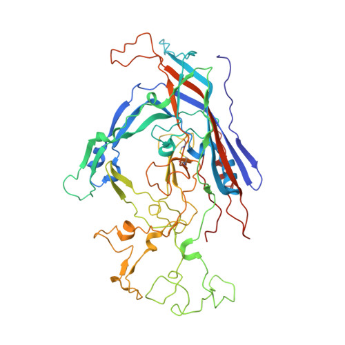
Canine parvovirus (CPV) emerged in 1978 as a host range variant of feline panleukopenia virus (FPV). This change of host was mediated by the mutation of five residues on the surface of the capsid. CPV and FPV enter cells by endocytosis and can be taken up by many non-permissive cell lines, showing that their host range and tissue specificity are largely determined by events occurring after cell entry. We have determined the structures of a variety of strains of CPV and FPV at various pH values and in the presence or absence of Ca(2+). The largest structural difference was found to occur in a flexible surface loop, consisting of residues 359 to 375 of the capsid protein. This loop binds a divalent calcium ion in FPV and is adjacent to a double Ca(2+)-binding site, both in CPV and FPV. Residues within the loop and those associated with the double Ca(2+)-binding site were found to be essential for virus infectivity. The residues involved in the double Ca(2+)-binding site are conserved only in FPV and CPV. Our results show that the loop conformation and the associated Ca(2+)-binding are influenced by the Ca(2+) concentration, as well as pH. These changes are correlated with the ability of the virus to hemagglutinate erythrocytes. The co-localization of hemagglutinating activity and host range determinants on the virus surface implies that these properties may be functionally linked. We speculate that the flexible loop and surrounding regions are involved in binding an as yet unidentified host molecule and that this interaction influences host range.
Organizational Affiliation:
Department of Biological Sciences, Purdue University, West Lafayette, IN, 47907-1392, USA.