1C4D

GRAMICIDIN CSCL COMPLEX


Experimental Data Snapshot

  • Method: X-RAY DIFFRACTION
  • Resolution: 2.00 Å
  • R-Value Work: 0.232 
  • R-Value Observed: 0.232 

wwPDB Validation   3D Report Full Report


This is version 2.0 of the entry. See complete history


Literature

The Gramicidin Pore: Crystal Structure of a Cesium Complex.

Wallace, B.A.Ravikumar, K.

(1988) Science 241: 182

  • DOI: https://doi.org/10.1126/science.2455344
  • Primary Citation of Related Structures:  
    1C4D

  • PubMed Abstract: 

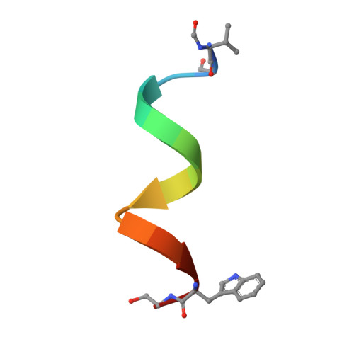
    Gramicidin, a linear polypeptide composed of hydrophobic amino acids with alternating L- and D- configurations, forms transmembrane ion channels. The crystal structure of a gramicidin-cesium complex has been determined at 2.0 angstrom resolution. In this structure, gramicidin forms a 26 angstrom long tube comprised of two polypeptide chains arranged as antiparallel beta strands that are wrapped into a left-handed helical coil with 6.4 residues per turn. The polypeptide backbone forms the interior of the hydrophilic, solvent-filled pore and the side chains form a hydrophobic and relatively regular surface on the outside of the pore. This example of a crystal structure of a solvent-filled ion pore provides a basis for understanding the physical nature of ion translocation.


  • Organizational Affiliation

    Department of Chemistry and Center for Biophysics, Rensselaer Polytechnic Institute, Troy, NY 12180.


Macromolecules

Find similar proteins by:  Sequence   |   3D Structure  

Entity ID: 1
MoleculeChains Sequence LengthOrganismDetailsImage
GRAMICIDIN A
A, B, C, D
16Brevibacillus brevisMutation(s): 0 
Entity Groups  
Sequence Clusters30% Identity50% Identity70% Identity90% Identity95% Identity100% Identity
Sequence Annotations
Expand
  • Reference Sequence
Small Molecules
Ligands 3 Unique
IDChains Name / Formula / InChI Key2D Diagram3D Interactions
CS
Query on CS

Download Ideal Coordinates CCD File 
E [auth A]
F [auth A]
G [auth A]
N [auth B]
S [auth C]
E [auth A],
F [auth A],
G [auth A],
N [auth B],
S [auth C],
X [auth D],
Y [auth D],
Z [auth D]
CESIUM ION
Cs
NCMHKCKGHRPLCM-UHFFFAOYSA-N
CL
Query on CL

Download Ideal Coordinates CCD File 
AA [auth D]
H [auth A]
I [auth A]
O [auth B]
T [auth C]
AA [auth D],
H [auth A],
I [auth A],
O [auth B],
T [auth C],
U [auth C]
CHLORIDE ION
Cl
VEXZGXHMUGYJMC-UHFFFAOYSA-M
MOH
Query on MOH

Download Ideal Coordinates CCD File 
BA [auth D]
CA [auth D]
DA [auth D]
EA [auth D]
J [auth A]
BA [auth D],
CA [auth D],
DA [auth D],
EA [auth D],
J [auth A],
K [auth A],
L [auth A],
M [auth A],
P [auth B],
Q [auth B],
R [auth B],
V [auth C],
W [auth C]
METHANOL
C H4 O
OKKJLVBELUTLKV-UHFFFAOYSA-N
Modified Residues  1 Unique
IDChains TypeFormula2D DiagramParent
FVA
Query on FVA
A, B, C, D
L-PEPTIDE LINKINGC6 H11 N O3VAL
Biologically Interesting Molecules (External Reference) 1 Unique
Experimental Data & Validation

Experimental Data

  • Method: X-RAY DIFFRACTION
  • Resolution: 2.00 Å
  • R-Value Work: 0.232 
  • R-Value Observed: 0.232 
  • Space Group: P 21 21 21
Unit Cell:
Length ( Å )Angle ( ˚ )
a = 32.118α = 90
b = 52.103β = 90
c = 31.174γ = 90
Software Package:
Software NamePurpose
PHASITmodel building
ASCALCmodel building
X-PLORrefinement
HANSONdata reduction
ANOSCLdata scaling
SCALE2data scaling
SCALE3data scaling
PHASITphasing
ASCALCphasing

Structure Validation

View Full Validation Report


Entry History 

Deposition Data

Revision History  (Full details and data files)

  • Version 1.0: 2000-01-03
    Type: Initial release
  • Version 1.1: 2011-06-14
    Changes: Version format compliance
  • Version 1.2: 2011-07-13
    Changes: Version format compliance
  • Version 1.3: 2011-07-27
    Changes: Atomic model, Database references, Derived calculations, Non-polymer description, Structure summary
  • Version 1.4: 2012-12-12
    Changes: Other
  • Version 1.5: 2017-10-04
    Changes: Refinement description
  • Version 2.0: 2023-11-15
    Changes: Atomic model, Data collection, Database references, Derived calculations