1B8D

CRYSTAL STRUCTURE OF A PHYCOUROBILIN-CONTAINING PHYCOERYTHRIN


Experimental Data Snapshot

  • Method: X-RAY DIFFRACTION
  • Resolution: 1.90 Å
  • R-Value Free: 0.227 
  • R-Value Work: 0.175 

wwPDB Validation   3D Report Full Report


Ligand Structure Quality Assessment 


This is version 1.3 of the entry. See complete history


Literature

Crystal structure of a phycourobilin-containing phycoerythrin at 1.90-A resolution.

Ritter, S.Hiller, R.G.Wrench, P.M.Welte, W.Diederichs, K.

(1999) J Struct Biol 126: 86-97

  • DOI: https://doi.org/10.1006/jsbi.1999.4106
  • Primary Citation of Related Structures:  
    1B8D

  • PubMed Abstract: 

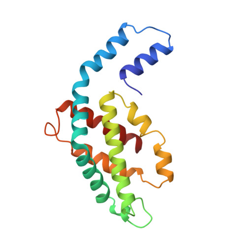
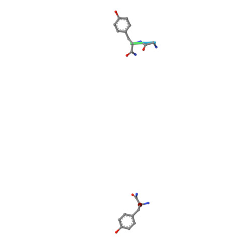
    The structure of R-phycoerythrin (R-PE) from the red alga Griffithsia monilis was solved at 1.90-A resolution by molecular replacement, using the atomic coordinates of cyanobacterial phycocyanin from Fremyella diplosiphon as a model. The crystallographic R factor for the final model is 17.5% (Rfree 22.7%) for reflections in the range 100-1.90 A. The model consists of an (alphabeta)2 dimer with an internal noncrystallographic dyad and a fragment of the gamma-polypeptide. The alpha-polypeptide (164 amino acid residues) has two covalently bound phycoerythrobilins at positions alpha82 and alpha139. The beta-polypeptide (177 residues) has two phycoerythrobilins bound to residues beta82 and beta158 and one phycourobilin covalently attached to rings A and D at residues beta50 and beta61, respectively. The electron density of the gamma-polypeptide is mostly averaged out by threefold crystallographic symmetry, but a dipeptide (Gly-Tyr) and one single Tyr could be modeled. These two tyrosine residues of the gamma-polypeptide are in close proximity to the phycoerythrobilins at position beta82 of two symmetry-related beta-polypeptides and are related by the same noncrystallographic dyad as the (alphabeta)2 dimer. Possible energy transfer pathways are discussed briefly.


  • Organizational Affiliation

    Institut für Biophysik und Strahlenbiologie, Universität Freiburg, Albertstrasse 23, Freiburg, D-79104, Germany.


Macromolecules
Find similar proteins by:  (by identity cutoff)  |  3D Structure
Entity ID: 1
MoleculeChains Sequence LengthOrganismDetailsImage
PROTEIN (RHODOPHYTAN PHYCOERYTHRIN (ALPHA CHAIN))A,
C [auth K]
164Griffithsia monilisMutation(s): 0 
UniProt
Find proteins for O36005 (Griffithsia monilis)
Explore O36005 
Go to UniProtKB:  O36005
Entity Groups  
Sequence Clusters30% Identity50% Identity70% Identity90% Identity95% Identity100% Identity
UniProt GroupO36005
Sequence Annotations
Expand
  • Reference Sequence
Find similar proteins by:  (by identity cutoff)  |  3D Structure
Entity ID: 2
MoleculeChains Sequence LengthOrganismDetailsImage
PROTEIN (RHODOPHYTAN PHYCOERYTHRIN (BETA CHAIN))B,
D [auth L]
177Griffithsia monilisMutation(s): 0 
UniProt
Find proteins for O36004 (Griffithsia monilis)
Explore O36004 
Go to UniProtKB:  O36004
Entity Groups  
Sequence Clusters30% Identity50% Identity70% Identity90% Identity95% Identity100% Identity
UniProt GroupO36004
Sequence Annotations
Expand
  • Reference Sequence

Find similar proteins by:  Sequence   |   3D Structure  

Entity ID: 3
MoleculeChains Sequence LengthOrganismDetailsImage
PROTEIN (RHODOPHYTAN PHYCOERYTHRIN (GAMMA CHAIN))E [auth G]6Griffithsia monilisMutation(s): 0 
Sequence Annotations
Expand
  • Reference Sequence
Small Molecules
Ligands 2 Unique
IDChains Name / Formula / InChI Key2D Diagram3D Interactions
PUB
Query on PUB

Download Ideal Coordinates CCD File 
J [auth B],
O [auth L]
PHYCOUROBILIN
C33 H42 N4 O6
KDCCOOGTVSRCHX-YYVBKQGDSA-N
PEB
Query on PEB

Download Ideal Coordinates CCD File 
F [auth A]
G [auth A]
H [auth B]
I [auth B]
K
F [auth A],
G [auth A],
H [auth B],
I [auth B],
K,
L [auth K],
M [auth L],
N [auth L]
PHYCOERYTHROBILIN
C33 H40 N4 O6
NKCBCVIFPXGHAV-WAVSMFBNSA-N
Modified Residues  1 Unique
IDChains TypeFormula2D DiagramParent
MEN
Query on MEN
B,
D [auth L]
L-PEPTIDE LINKINGC5 H10 N2 O3ASN
Experimental Data & Validation

Experimental Data

  • Method: X-RAY DIFFRACTION
  • Resolution: 1.90 Å
  • R-Value Free: 0.227 
  • R-Value Work: 0.175 
  • Space Group: H 3
Unit Cell:
Length ( Å )Angle ( ˚ )
a = 187.35α = 90
b = 187.35β = 90
c = 59.31γ = 120
Software Package:
Software NamePurpose
X-PLORmodel building
X-PLORrefinement
XDSdata reduction
XSCALEdata scaling
X-PLORphasing

Structure Validation

View Full Validation Report


Ligand Structure Quality Assessment 


Entry History 

Deposition Data

Revision History  (Full details and data files)

  • Version 1.0: 1999-02-18
    Type: Initial release
  • Version 1.1: 2008-04-26
    Changes: Version format compliance
  • Version 1.2: 2011-07-13
    Changes: Version format compliance
  • Version 1.3: 2023-08-09
    Changes: Data collection, Database references, Derived calculations, Refinement description