1ADW

PSEUDOAZURIN


Experimental Data Snapshot

  • Method: X-RAY DIFFRACTION
  • Resolution: 2.50 Å
  • R-Value Free: 0.239 
  • R-Value Work: 0.189 
  • R-Value Observed: 0.189 

wwPDB Validation   3D Report Full Report


This is version 1.3 of the entry. See complete history


Literature

Pseudospecific docking surfaces on electron transfer proteins as illustrated by pseudoazurin, cytochrome c550 and cytochrome cd1 nitrite reductase.

Williams, P.A.Fulop, V.Leung, Y.C.Chan, C.Moir, J.W.Howlett, G.Ferguson, S.J.Radford, S.E.Hajdu, J.

(1995) Nat Struct Biol 2: 975-982

  • DOI: https://doi.org/10.1038/nsb1195-975
  • Primary Citation of Related Structures:  
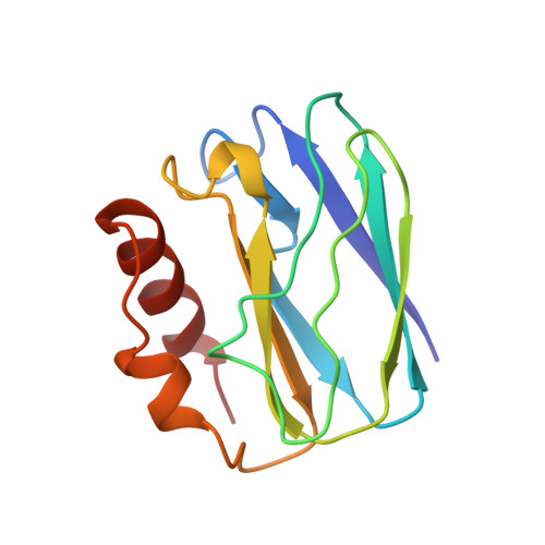
    1ADW

  • PubMed Abstract: 

    The structure of pseudoazurin from Thiosphaera pantotropha has been determined and compared to structures of both soluble and membrane-bound periplasmic redox proteins. The results show a matching set of unipolar, but promiscuous, docking motifs based on a positive hydrophobic surface patch on the electron shuttle proteins pseudoazurin and cytochrome c550 and a negative hydrophobic patch on the surface of their known redox partners. The observed electrostatic handedness is argued to be associated with the charge-asymmetry of the membrane-bound components of the redox chain due to von Heijne's 'positives-inside' principle. We propose a 'positives-in-between' rule for electron shuttle proteins, and expect a negative hydrophobic patch to be present on both the highest and lowest redox potential species in a series of electron carriers.


  • Organizational Affiliation

    Laboratory of Molecular Biophysics, University of Oxford, UK.


Macromolecules
Find similar proteins by:  (by identity cutoff)  |  3D Structure
Entity ID: 1
MoleculeChains Sequence LengthOrganismDetailsImage
PSEUDOAZURIN
A, B
123Paracoccus pantotrophusMutation(s): 0 
UniProt
Find proteins for P80401 (Paracoccus pantotrophus)
Explore P80401 
Go to UniProtKB:  P80401
Entity Groups  
Sequence Clusters30% Identity50% Identity70% Identity90% Identity95% Identity100% Identity
UniProt GroupP80401
Sequence Annotations
Expand
  • Reference Sequence
Experimental Data & Validation

Experimental Data

  • Method: X-RAY DIFFRACTION
  • Resolution: 2.50 Å
  • R-Value Free: 0.239 
  • R-Value Work: 0.189 
  • R-Value Observed: 0.189 
  • Space Group: C 1 2 1
Unit Cell:
Length ( Å )Angle ( ˚ )
a = 110α = 90
b = 58.4β = 127
c = 69.2γ = 90
Software Package:
Software NamePurpose
X-PLORmodel building
X-PLORrefinement
DENZOdata reduction
SCALEPACKdata scaling
X-PLORphasing

Structure Validation

View Full Validation Report


Entry History 

Deposition Data

Revision History  (Full details and data files)

  • Version 1.0: 1997-05-15
    Type: Initial release
  • Version 1.1: 2008-03-24
    Changes: Version format compliance
  • Version 1.2: 2011-07-13
    Changes: Version format compliance
  • Version 1.3: 2023-08-02
    Changes: Database references, Derived calculations, Other, Refinement description