1ZYO

Crystal Structure of the Serine Protease Domain of Sesbania Mosaic Virus polyprotein


Experimental Data Snapshot

  • Method: X-RAY DIFFRACTION
  • Resolution: 2.40 Å
  • R-Value Free: 0.288 
  • R-Value Work: 0.250 
  • R-Value Observed: 0.252 

wwPDB Validation   3D Report Full Report


This is version 1.2 of the entry. See complete history


Literature

Crystal structure of the serine protease domain of Sesbania mosaic virus polyprotein and mutational analysis of residues forming the S1-binding pocket

Gayathri, P.Satheshkumar, P.S.Prasad, K.Nair, S.Savithri, H.S.Murthy, M.R.N.

(2006) Virology 346: 440-451

  • DOI: https://doi.org/10.1016/j.virol.2005.11.011
  • Primary Citation of Related Structures:  
    1ZYO

  • PubMed Abstract: 

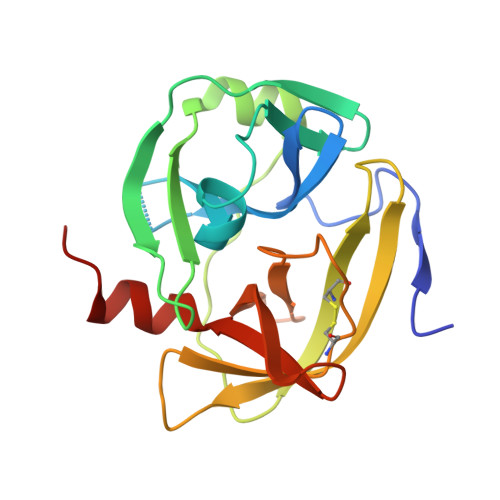
    Sesbania mosaic virus (SeMV) polyprotein is processed by its N-terminal serine protease domain. The crystal structure of the protease domain was determined to a resolution of 2.4 A using multiple isomorphous replacement and anomalous scattering. The SeMV protease domain exhibited the characteristic trypsin fold and was found to be closer to cellular serine proteases than to other viral proteases. The residues of the S1-binding pocket, H298, T279 and N308 were mutated to alanine in the DeltaN70-Protease-VPg polyprotein, and the cis-cleavage activity was examined. The H298A and T279A mutants were inactive, while the N308A mutant was partially active, suggesting that the interactions of H298 and T279 with P1-glutamate are crucial for the E-T/S cleavage. A region of exposed aromatic amino acids, probably essential for interaction with VPg, was identified on the protease domain, and this interaction could play a major role in modulating the function of the protease.


  • Organizational Affiliation

    Molecular Biophysics Unit, Indian Institute of Science, Bangalore 560 012, India.


Macromolecules
Find similar proteins by:  (by identity cutoff)  |  3D Structure
Entity ID: 1
MoleculeChains Sequence LengthOrganismDetailsImage
serine protease191Sesbania mosaic virusMutation(s): 0 
Gene Names: ORF2
EC: 3.4.21.19
UniProt
Find proteins for Q9EB08 (Sesbania mosaic virus)
Explore Q9EB08 
Go to UniProtKB:  Q9EB08
Entity Groups  
Sequence Clusters30% Identity50% Identity70% Identity90% Identity95% Identity100% Identity
UniProt GroupQ9EB08
Sequence Annotations
Expand
  • Reference Sequence
Small Molecules
Ligands 1 Unique
IDChains Name / Formula / InChI Key2D Diagram3D Interactions
GOL
Query on GOL

Download Ideal Coordinates CCD File 
B [auth A]GLYCEROL
C3 H8 O3
PEDCQBHIVMGVHV-UHFFFAOYSA-N
Experimental Data & Validation

Experimental Data

  • Method: X-RAY DIFFRACTION
  • Resolution: 2.40 Å
  • R-Value Free: 0.288 
  • R-Value Work: 0.250 
  • R-Value Observed: 0.252 
  • Space Group: P 31 2 1
Unit Cell:
Length ( Å )Angle ( ˚ )
a = 74.072α = 90
b = 74.072β = 90
c = 68.753γ = 120
Software Package:
Software NamePurpose
REFMACrefinement
DENZOdata reduction
SCALEPACKdata scaling
MLPHAREphasing

Structure Validation

View Full Validation Report


Entry History 

Deposition Data

Revision History  (Full details and data files)

  • Version 1.0: 2006-04-25
    Type: Initial release
  • Version 1.1: 2008-04-30
    Changes: Version format compliance
  • Version 1.2: 2011-07-13
    Changes: Non-polymer description, Version format compliance