1ZVZ

Vinculin Head (0-258) in Complex with the Talin Rod Residue 820-844


Experimental Data Snapshot

  • Method: X-RAY DIFFRACTION
  • Resolution: 1.80 Å
  • R-Value Free: 0.276 
  • R-Value Work: 0.239 
  • R-Value Observed: 0.241 

wwPDB Validation   3D Report Full Report


This is version 1.3 of the entry. See complete history


Literature

Mapping and consensus sequence identification for multiple vinculin binding sites within the talin rod

Gingras, A.R.Ziegler, W.H.Frank, R.Barsukov, I.L.Roberts, G.C.Critchley, D.R.Emsley, J.

(2005) J Biol Chem 280: 37217-37224

  • DOI: https://doi.org/10.1074/jbc.M508060200
  • Primary Citation of Related Structures:  
    1ZVZ, 1ZW2, 1ZW3

  • PubMed Abstract: 

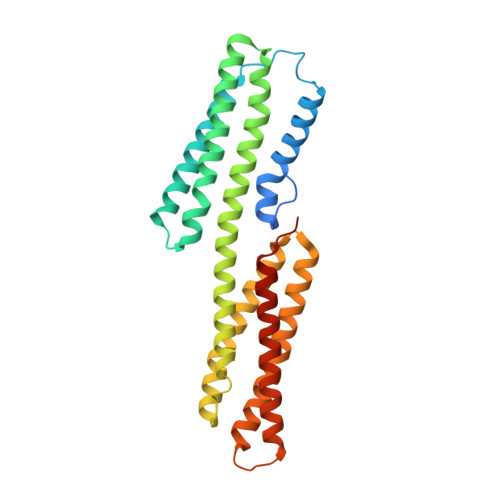
    The interaction between the cytoskeletal proteins talin and vinculin plays a key role in integrin-mediated cell adhesion and migration. Three vinculin binding sites (VBS1-3) have previously been identified in the talin rod using a yeast two-hybrid assay. To extend these studies, we spot-synthesized a series of peptides spanning all the alpha-helical regions predicted for the talin rod and identified eight additional VBSs, two of which overlap key functional regions of the rod, including the integrin binding site and C-terminal actin binding site. The talin VBS alpha-helices bind to a hydrophobic cleft in the N-terminal vinculin Vd1 domain. We have defined the specificity of this interaction by spot-synthesizing a series of 25-mer talin VBS1 peptides containing substitutions with all the commonly occurring amino acids. The consensus for recognition is LXXAAXXVAXX- VXXLIXXA with distinct classes of hydrophobic side chains at positions 1, 4, 5, 8, 9, 12, 15, and 16 required for vinculin binding. Positions 1, 8, 12, 15, and 16 require an aliphatic residue and will not tolerate alanine, whereas positions 4, 5, and 9 are less restrictive. These preferences are common to all 11 VBS sequences with a minor variation occurring in one case. A crystal structure of this variant VBS peptide in complex with the vinculin Vd1 domain reveals a subtly different mode of vinculin binding.


  • Organizational Affiliation

    Department of Biochemistry, University of Leicester, Leicester LE1 7RH, United Kingdom.


Macromolecules
Find similar proteins by:  (by identity cutoff)  |  3D Structure
Entity ID: 1
MoleculeChains Sequence LengthOrganismDetailsImage
Vinculin279Gallus gallusMutation(s): 0 
Gene Names: VCLVINC1
UniProt
Find proteins for P12003 (Gallus gallus)
Explore P12003 
Go to UniProtKB:  P12003
Entity Groups  
Sequence Clusters30% Identity50% Identity70% Identity90% Identity95% Identity100% Identity
UniProt GroupP12003
Sequence Annotations
Expand
  • Reference Sequence

Find similar proteins by:  Sequence   |   3D Structure  

Entity ID: 2
MoleculeChains Sequence LengthOrganismDetailsImage
Talin 125N/AMutation(s): 0 
UniProt
Find proteins for P54939 (Gallus gallus)
Explore P54939 
Go to UniProtKB:  P54939
Entity Groups  
Sequence Clusters30% Identity50% Identity70% Identity90% Identity95% Identity100% Identity
UniProt GroupP54939
Sequence Annotations
Expand
  • Reference Sequence
Experimental Data & Validation

Experimental Data

  • Method: X-RAY DIFFRACTION
  • Resolution: 1.80 Å
  • R-Value Free: 0.276 
  • R-Value Work: 0.239 
  • R-Value Observed: 0.241 
  • Space Group: P 21 21 21
Unit Cell:
Length ( Å )Angle ( ˚ )
a = 36.013α = 90
b = 52.164β = 90
c = 153.841γ = 90
Software Package:
Software NamePurpose
REFMACrefinement
DENZOdata reduction
SCALEPACKdata scaling
PHASERphasing

Structure Validation

View Full Validation Report


Entry History 

Deposition Data

Revision History  (Full details and data files)

  • Version 1.0: 2005-09-20
    Type: Initial release
  • Version 1.1: 2008-04-30
    Changes: Version format compliance
  • Version 1.2: 2011-07-13
    Changes: Version format compliance
  • Version 1.3: 2023-08-23
    Changes: Data collection, Database references, Refinement description