1ZRZ

Crystal Structure of the Catalytic Domain of Atypical Protein Kinase C-iota


Experimental Data Snapshot

  • Method: X-RAY DIFFRACTION
  • Resolution: 3.00 Å
  • R-Value Free: 0.333 
  • R-Value Work: 0.249 
  • R-Value Observed: 0.249 

wwPDB Validation   3D Report Full Report


Ligand Structure Quality Assessment 


This is version 1.3 of the entry. See complete history


Literature

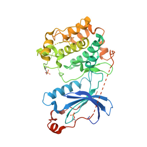
Crystal Structure of the Catalytic Domain of Human Atypical Protein Kinase C-iota Reveals Interaction Mode of Phosphorylation Site in Turn Motif

Messerschmidt, A.Macieira, S.Velarde, M.Baedeker, M.Benda, C.Jestel, A.Brandstetter, H.Neuefeind, T.Blaesse, M.

(2005) J Mol Biol 352: 918-931

  • DOI: https://doi.org/10.1016/j.jmb.2005.07.060
  • Primary Citation of Related Structures:  
    1ZRZ

  • PubMed Abstract: 

    Atypical protein kinases C (aPKCs) play critical roles in signaling pathways that control cell growth, differentiation and survival. Therefore, they constitute attractive targets for the development of novel therapeutics against cancer. The crystal structure of the catalytic domain of atypical PKCiota in complex with the bis(indolyl)maleimide inhibitor BIM1 has been determined at 3.0A resolution within the frame of the European Structural Proteomics Project SPINE. The overall structure exhibits the classical bilobal kinase fold and is in its fully activated form. Both phosphorylation sites (Thr403 in the activation loop, and Thr555 in the turn motif) are well defined in the structure and form intramolecular ionic contacts that make an important contribution in stabilizing the active conformation of the catalytic subunit. The phosphorylation site in the hydrophobic motif of atypical PKCs is replaced by the phosphorylation mimic glutamate and this is also clearly seen in the structure of PKCiota (residue 574). This structure determination for the first time provides the architecture of the turn motif phosphorylation site, which is characteristic for PKCs and PKB/AKT, and is completely different from that in PKA. The bound BIM1 inhibitor blocks the ATP-binding site and puts the kinase domain into an intermediate open conformation. The PKCiota-BIM1 complex is the first kinase domain crystal structure of any atypical PKC and constitutes the basis for rational drug design for selective PKCiota inhibitors.


  • Organizational Affiliation

    Department of Structural Research, Max-Planck-Institute of Biochemistry, Am Klopferspitz 18, 82152 Martinsried, Germany. messersc@biochem.mpg.de


Macromolecules
Find similar proteins by:  (by identity cutoff)  |  3D Structure
Entity ID: 1
MoleculeChains Sequence LengthOrganismDetailsImage
Protein kinase C, iota364Homo sapiensMutation(s): 2 
Gene Names: PRKCI
EC: 2.7.1
UniProt & NIH Common Fund Data Resources
Find proteins for P41743 (Homo sapiens)
Explore P41743 
Go to UniProtKB:  P41743
PHAROS:  P41743
GTEx:  ENSG00000163558 
Entity Groups  
Sequence Clusters30% Identity50% Identity70% Identity90% Identity95% Identity100% Identity
UniProt GroupP41743
Sequence Annotations
Expand
  • Reference Sequence
Small Molecules
Ligands 1 Unique
IDChains Name / Formula / InChI Key2D Diagram3D Interactions
BI1
Query on BI1

Download Ideal Coordinates CCD File 
B [auth A]3-{1-[3-(DIMETHYLAMINO)PROPYL]-1H-INDOL-3-YL}-4-(1H-INDOL-3-YL)-1H-PYRROLE-2,5-DIONE
C25 H24 N4 O2
QMGUOJYZJKLOLH-UHFFFAOYSA-N
Modified Residues  1 Unique
IDChains TypeFormula2D DiagramParent
TPO
Query on TPO
A
L-PEPTIDE LINKINGC4 H10 N O6 PTHR
Binding Affinity Annotations 
IDSourceBinding Affinity
BI1 BindingDB:  1ZRZ IC50: 3463 (nM) from 1 assay(s)
PDBBind:  1ZRZ IC50: 5800 (nM) from 1 assay(s)
Experimental Data & Validation

Experimental Data

  • Method: X-RAY DIFFRACTION
  • Resolution: 3.00 Å
  • R-Value Free: 0.333 
  • R-Value Work: 0.249 
  • R-Value Observed: 0.249 
  • Space Group: P 32 2 1
Unit Cell:
Length ( Å )Angle ( ˚ )
a = 78.143α = 90
b = 78.143β = 90
c = 112.625γ = 120
Software Package:
Software NamePurpose
CNSrefinement
MOSFLMdata reduction
CCP4data scaling
MOLREPphasing

Structure Validation

View Full Validation Report


Ligand Structure Quality Assessment 


Entry History 

Revision History  (Full details and data files)

  • Version 1.0: 2005-09-13
    Type: Initial release
  • Version 1.1: 2008-04-30
    Changes: Version format compliance
  • Version 1.2: 2011-07-13
    Changes: Version format compliance
  • Version 1.3: 2023-08-23
    Changes: Data collection, Database references, Derived calculations, Refinement description