1Z59

Topoisomerase VI-B, ADP-bound monomer form


Experimental Data Snapshot

  • Method: X-RAY DIFFRACTION
  • Resolution: 2.10 Å
  • R-Value Free: 0.228 
  • R-Value Work: 0.189 
  • R-Value Observed: 0.192 

wwPDB Validation   3D Report Full Report


Ligand Structure Quality Assessment 


This is version 1.4 of the entry. See complete history


Literature

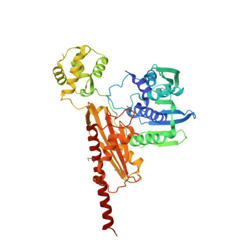
Structural dissection of ATP turnover in the prototypical GHL ATPase TopoVI.

Corbett, K.D.Berger, J.M.

(2005) Structure 13: 873-882

  • DOI: https://doi.org/10.1016/j.str.2005.03.013
  • Primary Citation of Related Structures:  
    1Z59, 1Z5A, 1Z5B, 1Z5C

  • PubMed Abstract: 

    GHL proteins are functionally diverse enzymes defined by the presence of a conserved ATPase domain that self-associates to trap substrate upon nucleotide binding. The structural states adopted by these enzymes during nucleotide hydrolysis and product release, and their consequences for enzyme catalysis, have remained unclear. Here, we have determined a complete structural map of the ATP turnover cycle for topoVI-B, the ATPase subunit of the archaeal GHL enzyme topoisomerase VI. With this ensemble of structures, we show that significant conformational changes in the subunit occur first upon ATP binding, and subsequently upon release of hydrolyzed P(i). Together, these data provide a structural framework for understanding the role of ATP hydrolysis in the type II topoisomerase reaction. Our results also suggest that the GHL ATPase module is a molecular switch in which ATP hydrolysis serves as a prerequisite but not a driving force for substrate-dependent structural transitions in the enzyme.


  • Organizational Affiliation

    Department of Molecular and cellular biology, University of California, Berkeley, California 94720, USA.


Macromolecules
Find similar proteins by:  (by identity cutoff)  |  3D Structure
Entity ID: 1
MoleculeChains Sequence LengthOrganismDetailsImage
Type II DNA topoisomerase VI subunit B469Saccharolobus shibataeMutation(s): 5 
Gene Names: top6B
EC: 5.99.1.3
UniProt
Find proteins for O05207 (Saccharolobus shibatae (strain ATCC 51178 / DSM 5389 / JCM 8931 / NBRC 15437 / B12))
Explore O05207 
Go to UniProtKB:  O05207
Entity Groups  
Sequence Clusters30% Identity50% Identity70% Identity90% Identity95% Identity100% Identity
UniProt GroupO05207
Sequence Annotations
Expand
  • Reference Sequence
Small Molecules
Ligands 2 Unique
IDChains Name / Formula / InChI Key2D Diagram3D Interactions
ADP
Query on ADP

Download Ideal Coordinates CCD File 
C [auth A]ADENOSINE-5'-DIPHOSPHATE
C10 H15 N5 O10 P2
XTWYTFMLZFPYCI-KQYNXXCUSA-N
MG
Query on MG

Download Ideal Coordinates CCD File 
B [auth A]MAGNESIUM ION
Mg
JLVVSXFLKOJNIY-UHFFFAOYSA-N
Modified Residues  1 Unique
IDChains TypeFormula2D DiagramParent
MSE
Query on MSE
A
L-PEPTIDE LINKINGC5 H11 N O2 SeMET
Experimental Data & Validation

Experimental Data

  • Method: X-RAY DIFFRACTION
  • Resolution: 2.10 Å
  • R-Value Free: 0.228 
  • R-Value Work: 0.189 
  • R-Value Observed: 0.192 
  • Space Group: P 21 21 2
Unit Cell:
Length ( Å )Angle ( ˚ )
a = 94.573α = 90
b = 112.451β = 90
c = 56.079γ = 90
Software Package:
Software NamePurpose
REFMACrefinement
HKL-2000data reduction
SCALEPACKdata scaling
AMoREphasing

Structure Validation

View Full Validation Report


Ligand Structure Quality Assessment 


Entry History 

Deposition Data

Revision History  (Full details and data files)

  • Version 1.0: 2005-06-14
    Type: Initial release
  • Version 1.1: 2008-04-30
    Changes: Version format compliance
  • Version 1.2: 2011-07-13
    Changes: Advisory, Version format compliance
  • Version 1.3: 2023-08-23
    Changes: Data collection, Database references, Derived calculations, Refinement description
  • Version 1.4: 2023-11-15
    Changes: Data collection