1YRX

Structure of a novel photoreceptor: the BLUF domain of AppA from Rhodobacter sphaeroides


Experimental Data Snapshot

  • Method: X-RAY DIFFRACTION
  • Resolution: 2.30 Å
  • R-Value Free: 0.273 
  • R-Value Work: 0.241 
  • R-Value Observed: 0.243 

wwPDB Validation   3D Report Full Report


This is version 1.3 of the entry. See complete history


Literature

Structure of a Novel Photoreceptor, the BLUF Domain of AppA from Rhodobacter sphaeroides

Anderson, S.Dragnea, V.Masuda, S.Ybe, J.Moffat, K.Bauer, C.

(2005) Biochemistry 44: 7998-8005

  • DOI: https://doi.org/10.1021/bi0502691
  • Primary Citation of Related Structures:  
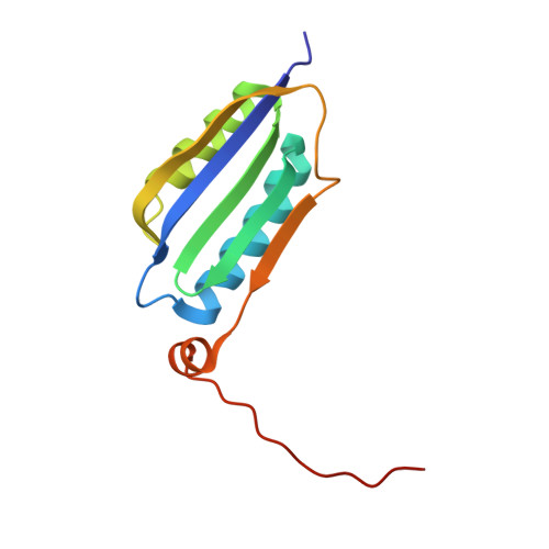
    1YRX

  • PubMed Abstract: 

    The flavin-binding BLUF domain of AppA represents a new class of blue light photoreceptors that are present in a number of bacterial and algal species. The dark state X-ray structure of this domain was determined at 2.3 A resolution. The domain demonstrates a new function for the common ferredoxin-like fold; two long alpha-helices flank the flavin, which is bound with its isoalloxazine ring perpendicular to a five-stranded beta-sheet. The hydrogen bond network and the overall protein topology of the BLUF domain (but not its sequence) bear some resemblance to LOV domains, a subset of PAS domains widely involved in signaling. Nearly all residues conserved in BLUF domains surround the flavin chromophore, many of which are involved in an intricate hydrogen bond network. Photoactivation may induce a rearrangement in this network via reorientation of the Gln63 side chain to form a new hydrogen bond to the flavin O4 position. This shift would also break a hydrogen bond to the Trp104 side chain, which may be critical in induction of global structural change in AppA.


  • Organizational Affiliation

    Consortium for Advanced Radiation Sources, Department of Biochemistry and Molecular Biology, University of Chicago, Chicago, Illinois 60637, USA. smander@uchicago.edu


Macromolecules
Find similar proteins by:  (by identity cutoff)  |  3D Structure
Entity ID: 1
MoleculeChains Sequence LengthOrganismDetailsImage
hypothetical protein Rsph03001874
A, B, C
121Cereibacter sphaeroides 2.4.1Mutation(s): 0 
UniProt
Find proteins for Q53119 (Cereibacter sphaeroides)
Explore Q53119 
Go to UniProtKB:  Q53119
Entity Groups  
Sequence Clusters30% Identity50% Identity70% Identity90% Identity95% Identity100% Identity
UniProt GroupQ53119
Sequence Annotations
Expand
  • Reference Sequence
Small Molecules
Ligands 2 Unique
IDChains Name / Formula / InChI Key2D Diagram3D Interactions
FMN
Query on FMN

Download Ideal Coordinates CCD File 
D [auth A],
H [auth B],
I [auth C]
FLAVIN MONONUCLEOTIDE
C17 H21 N4 O9 P
FVTCRASFADXXNN-SCRDCRAPSA-N
D9G
Query on D9G

Download Ideal Coordinates CCD File 
E [auth A]
F [auth A]
G [auth A]
J [auth C]
K [auth C]
E [auth A],
F [auth A],
G [auth A],
J [auth C],
K [auth C],
L [auth C]
N-DODECYL-N,N-DIMETHYLGLYCINATE
C16 H33 N O2
DVEKCXOJTLDBFE-UHFFFAOYSA-N
Experimental Data & Validation

Experimental Data

  • Method: X-RAY DIFFRACTION
  • Resolution: 2.30 Å
  • R-Value Free: 0.273 
  • R-Value Work: 0.241 
  • R-Value Observed: 0.243 
  • Space Group: P 41 21 2
Unit Cell:
Length ( Å )Angle ( ˚ )
a = 98.671α = 90
b = 98.671β = 90
c = 127.453γ = 90
Software Package:
Software NamePurpose
HKL-2000data collection
SCALEPACKdata scaling
XTALVIEWrefinement
REFMACrefinement
HKL-2000data reduction

Structure Validation

View Full Validation Report


Entry History 

Deposition Data

Revision History  (Full details and data files)

  • Version 1.0: 2005-06-28
    Type: Initial release
  • Version 1.1: 2008-04-30
    Changes: Version format compliance
  • Version 1.2: 2011-07-13
    Changes: Derived calculations, Version format compliance
  • Version 1.3: 2024-02-14
    Changes: Data collection, Database references, Derived calculations