1YHU

Crystal structure of Riftia pachyptila C1 hemoglobin reveals novel assembly of 24 subunits.


Experimental Data Snapshot

  • Method: X-RAY DIFFRACTION
  • Resolution: 3.15 Å
  • R-Value Free: 0.271 
  • R-Value Work: 0.243 
  • R-Value Observed: 0.243 

wwPDB Validation   3D Report Full Report


Ligand Structure Quality Assessment 


This is version 1.3 of the entry. See complete history


Literature

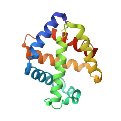
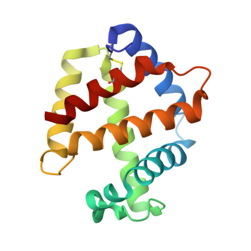
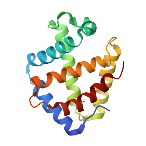
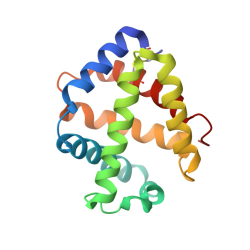
Sulfide binding is mediated by zinc ions discovered in the crystal structure of a hydrothermal vent tubeworm hemoglobin.

Flores, J.F.Fisher, C.R.Carney, S.L.Green, B.N.Freytag, J.K.Schaeffer, S.W.Royer Jr, W.E.

(2005) Proc Natl Acad Sci U S A 102: 2713-2718

  • DOI: https://doi.org/10.1073/pnas.0407455102
  • Primary Citation of Related Structures:  
    1YHU

  • PubMed Abstract: 

    Key to the remarkable ability of vestimentiferan tubeworms to thrive in the harsh conditions of hydrothermal vents are hemoglobins that permit the sequestration and delivery of hydrogen sulfide and oxygen to chemoautotrophic bacteria. Here, we demonstrate that zinc ions, not free cysteine residues, bind sulfide in vestimentiferan hemoglobins. The crystal structure of the C1 hemoglobin from the hydrothermal vent tubeworm Riftia pachyptila has been determined to 3.15 A and revealed the unexpected presence of 12 tightly bound Zn(2+) ions near the threefold axes of this D(3) symmetric hollow sphere. Chelation experiments on R. pachyptila whole-coelomic fluid and purified hemoglobins reveal a role for Zn(2+) ions in sulfide binding. Free cysteine residues, previously proposed as sulfide-binding sites in vestimentiferan hemoglobins, are found buried in surprisingly hydrophobic pockets below the surface of the R. pachyptila C1 molecule, suggesting that access of these residues to environmental sulfide is restricted. Attempts to reduce the sulfide-binding capacities of R. pachyptila hemoglobins by addition of a thiol inhibitor were also unsuccessful. These findings challenge the currently accepted paradigm of annelid hemoglobin evolution and adaptation to reducing environments.


  • Organizational Affiliation

    Department of Biology, Pennsylvania State University, University Park, PA 16802, USA. jff133@psu.edu


Macromolecules
Find similar proteins by:  (by identity cutoff)  |  3D Structure
Entity ID: 1
MoleculeChains Sequence LengthOrganismDetailsImage
hemoglobin A1 chain
A, E, I, M, Q
A, E, I, M, Q, U
145Riftia pachyptilaMutation(s): 0 
UniProt
Find proteins for Q8IFK4 (Riftia pachyptila)
Explore Q8IFK4 
Go to UniProtKB:  Q8IFK4
Entity Groups  
Sequence Clusters30% Identity50% Identity70% Identity90% Identity95% Identity100% Identity
UniProt GroupQ8IFK4
Sequence Annotations
Expand
  • Reference Sequence
Find similar proteins by:  (by identity cutoff)  |  3D Structure
Entity ID: 2
MoleculeChains Sequence LengthOrganismDetailsImage
Giant hemoglobins B chain
B, F, J, N, R
B, F, J, N, R, V
144Riftia pachyptilaMutation(s): 0 
UniProt
Find proteins for P80592 (Riftia pachyptila)
Explore P80592 
Go to UniProtKB:  P80592
Entity Groups  
Sequence Clusters30% Identity50% Identity70% Identity90% Identity95% Identity100% Identity
UniProt GroupP80592
Sequence Annotations
Expand
  • Reference Sequence
Find similar proteins by:  (by identity cutoff)  |  3D Structure
Entity ID: 3
MoleculeChains Sequence LengthOrganismDetailsImage
hemoglobin B1a chain
C, G, K, O, S
C, G, K, O, S, W
148Riftia pachyptilaMutation(s): 0 
UniProt
Find proteins for Q8IFK2 (Riftia pachyptila)
Explore Q8IFK2 
Go to UniProtKB:  Q8IFK2
Entity Groups  
Sequence Clusters30% Identity50% Identity70% Identity90% Identity95% Identity100% Identity
UniProt GroupQ8IFK2
Sequence Annotations
Expand
  • Reference Sequence
Find similar proteins by:  (by identity cutoff)  |  3D Structure
Entity ID: 4
MoleculeChains Sequence LengthOrganismDetailsImage
hemoglobin B2 chain
D, H, L, P, T
D, H, L, P, T, X
149Riftia pachyptilaMutation(s): 0 
UniProt
Find proteins for Q8IFJ9 (Riftia pachyptila)
Explore Q8IFJ9 
Go to UniProtKB:  Q8IFJ9
Entity Groups  
Sequence Clusters30% Identity50% Identity70% Identity90% Identity95% Identity100% Identity
UniProt GroupQ8IFJ9
Sequence Annotations
Expand
  • Reference Sequence
Small Molecules
Ligands 3 Unique
IDChains Name / Formula / InChI Key2D Diagram3D Interactions
HEM
Query on HEM

Download Ideal Coordinates CCD File 
AB [auth L]
AC [auth V]
CA [auth B]
CB [auth M]
CC [auth W]
AB [auth L],
AC [auth V],
CA [auth B],
CB [auth M],
CC [auth W],
EA [auth C],
EC [auth X],
GA [auth D],
GB [auth N],
IA [auth E],
IB [auth O],
KB [auth P],
MA [auth F],
MB [auth Q],
OA [auth G],
QA [auth H],
QB [auth R],
SA [auth I],
SB [auth S],
UB [auth T],
WA [auth J],
WB [auth U],
Y [auth A],
YA [auth K]
PROTOPORPHYRIN IX CONTAINING FE
C34 H32 Fe N4 O4
KABFMIBPWCXCRK-RGGAHWMASA-L
ZN
Query on ZN

Download Ideal Coordinates CCD File 
AA [auth B]
BA [auth B]
EB [auth N]
FB [auth N]
KA [auth F]
AA [auth B],
BA [auth B],
EB [auth N],
FB [auth N],
KA [auth F],
LA [auth F],
OB [auth R],
PB [auth R],
UA [auth J],
VA [auth J],
YB [auth V],
ZB [auth V]
ZINC ION
Zn
PTFCDOFLOPIGGS-UHFFFAOYSA-N
OXY
Query on OXY

Download Ideal Coordinates CCD File 
BB [auth L]
BC [auth V]
DA [auth B]
DB [auth M]
DC [auth W]
BB [auth L],
BC [auth V],
DA [auth B],
DB [auth M],
DC [auth W],
FA [auth C],
FC [auth X],
HA [auth D],
HB [auth N],
JA [auth E],
JB [auth O],
LB [auth P],
NA [auth F],
NB [auth Q],
PA [auth G],
RA [auth H],
RB [auth R],
TA [auth I],
TB [auth S],
VB [auth T],
XA [auth J],
XB [auth U],
Z [auth A],
ZA [auth K]
OXYGEN MOLECULE
O2
MYMOFIZGZYHOMD-UHFFFAOYSA-N
Experimental Data & Validation

Experimental Data

  • Method: X-RAY DIFFRACTION
  • Resolution: 3.15 Å
  • R-Value Free: 0.271 
  • R-Value Work: 0.243 
  • R-Value Observed: 0.243 
  • Space Group: P 41 21 2
Unit Cell:
Length ( Å )Angle ( ˚ )
a = 195.84α = 90
b = 195.84β = 90
c = 308.82γ = 90
Software Package:
Software NamePurpose
DENZOdata reduction
SCALEPACKdata scaling
CNSrefinement
CNSphasing

Structure Validation

View Full Validation Report


Ligand Structure Quality Assessment 


Entry History 

Deposition Data

Revision History  (Full details and data files)

  • Version 1.0: 2005-02-08
    Type: Initial release
  • Version 1.1: 2008-04-01
    Changes: Version format compliance
  • Version 1.2: 2011-07-13
    Changes: Derived calculations, Version format compliance
  • Version 1.3: 2013-03-06
    Changes: Data collection