Crystal structures of native and recombinant yeast fumarase.
Weaver, T., Lees, M., Zaitsev, V., Zaitseva, I., Duke, E., Lindley, P., McSweeny, S., Svensson, A., Keruchenko, J., Keruchenko, I., Gladilin, K., Banaszak, L.(1998) J Mol Biol 280: 431-442
- PubMed: 9665847
- DOI: https://doi.org/10.1006/jmbi.1998.1862
- Primary Citation of Related Structures:
1YFM - PubMed Abstract:
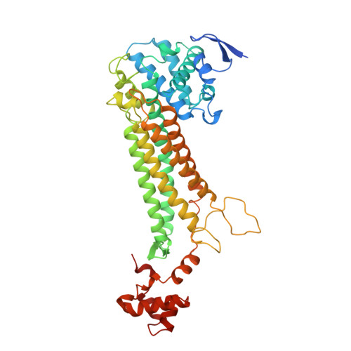
Crystal structures for both native and recombinant forms of yeast fumarase from Saccharomyces cerevisiae have been completed to moderate resolution by two separate laboratories. The recombinant form was obtained by the construction of an expression plasmid for Escherichia coli. Despite a high level of amino acid sequence similarity, purification of the eukaryotic enzyme from the wild-type prokaryotic enzyme was feasible. The crystal structure of the native form, NY-fumarase, encompasses residues R22 through M484, while the recombinant form, RY-fumarase, consists of residues S27 through L485. Both crystal structures lack the N-terminal translocation segment. Each subunit of the homo-tetrameric protein has three domains. The active site is formed by segments from each of three polypeptide chains. The results of these studies on the eukaryotic proteins are unique, since the recombinant form was done in the absence of dicarboxylic acid and has an unoccupied active site. As a comparison, native fumarase was crystallized in the presence of the competitive inhibitor, meso-tartrate. Meso-tartrate occupies a position close to that of the bound citrate molecule found in the active site of the E. coli enzyme. This inhibitor participates in hydrogen bonding to an active-site water molecule. The independent determination of the two structures provides further evidence that an active-site water molecule may play an active role in the fumarase-catalyzed reaction.
Organizational Affiliation:
Department of Biochemistry, University of Minnesota, 4-225 Millard Hall, Minneapolis, MN 55455-0347, USA.