1YE9

Crystal structure of proteolytically truncated catalase HPII from E. coli


Experimental Data Snapshot

  • Method: X-RAY DIFFRACTION
  • Resolution: 2.80 Å
  • R-Value Free: 0.269 
  • R-Value Work: 0.217 
  • R-Value Observed: 0.220 

wwPDB Validation   3D Report Full Report


Ligand Structure Quality Assessment 


This is version 1.3 of the entry. See complete history


Literature

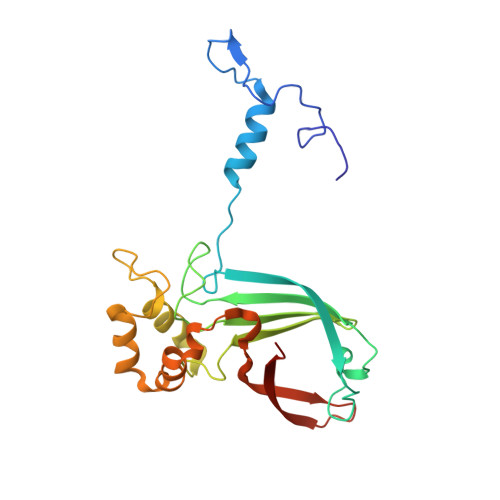
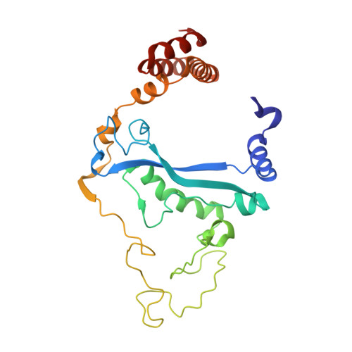
Characterization of a Large Subunit Catalase Truncated by Proteolytic Cleavage(,)

Chelikani, P.Carpena, X.Perez-Luque, R.Donald, L.J.Duckworth, H.W.Switala, J.Fita, I.Loewen, P.C.

(2005) Biochemistry 44: 5597-5605

  • DOI: https://doi.org/10.1021/bi047277m
  • Primary Citation of Related Structures:  
    1YE9

  • PubMed Abstract: 

    The large subunit catalase HPII from Escherichia coli can be truncated by proteolysis to a structure similar to small subunit catalases. Mass spectrometry analysis indicates that there is some heterogeneity in the precise cleavage sites, but approximately 74 N-terminal residues, 189 C-terminal residues, and a 9-11-residue internal fragment, including residues 298-308, are removed. Crystal structure refinement at 2.8 A reveals that the tertiary and quaternary structure of the native enzyme is retained with only very subtle changes despite the loss of 36% of the sequence. The truncated variant exhibits a 1.8 times faster turnover rate and enhanced sensitivity to high concentrations of H(2)O(2), consistent with easier access of the substrate to the active site. In addition, the truncated variant is more sensitive to inhibition, particularly by reagents such as aminotriazole and azide which are larger than substrate H(2)O(2). The main channel leading to the heme cavity is largely unaffected by the truncation, but the lateral channel is shortened and its entrance widened by removal of the C-terminal domain, providing an explanation for easier access to the active site. Opening of the entrance to the lateral channel also opens the putative NADPH binding site, but NADPH binding could not be demonstrated. Despite the lack of bound NADPH, the compound I species of both native and truncated HPII are reduced back to the resting state with compound II being evident in the absorbance spectrum only of the heme b-containing H392A variant.


  • Organizational Affiliation

    Department of Microbiology, University of Manitoba, Winnipeg, Manitoba R3T 2N2, Canada.


Macromolecules
Find similar proteins by:  (by identity cutoff)  |  3D Structure
Entity ID: 1
MoleculeChains Sequence LengthOrganismDetailsImage
catalase HPII226Escherichia coliMutation(s): 0 
Gene Names: KATE
EC: 1.11.1.6
UniProt
Find proteins for P21179 (Escherichia coli (strain K12))
Explore P21179 
Go to UniProtKB:  P21179
Entity Groups  
Sequence Clusters30% Identity50% Identity70% Identity90% Identity95% Identity100% Identity
UniProt GroupP21179
Sequence Annotations
Expand
  • Reference Sequence
Find similar proteins by:  (by identity cutoff)  |  3D Structure
Entity ID: 2
MoleculeChains Sequence LengthOrganismDetailsImage
catalase HPII259Escherichia coliMutation(s): 0 
Gene Names: KATE
EC: 1.11.1.6
UniProt
Find proteins for P21179 (Escherichia coli (strain K12))
Explore P21179 
Go to UniProtKB:  P21179
Entity Groups  
Sequence Clusters30% Identity50% Identity70% Identity90% Identity95% Identity100% Identity
UniProt GroupP21179
Sequence Annotations
Expand
  • Reference Sequence
Experimental Data & Validation

Experimental Data

  • Method: X-RAY DIFFRACTION
  • Resolution: 2.80 Å
  • R-Value Free: 0.269 
  • R-Value Work: 0.217 
  • R-Value Observed: 0.220 
  • Space Group: P 1 21 1
Unit Cell:
Length ( Å )Angle ( ˚ )
a = 111.011α = 90
b = 152.888β = 97.54
c = 135.287γ = 90
Software Package:
Software NamePurpose
REFMACrefinement
MOSFLMdata reduction
CCP4data scaling
MOLREPphasing

Structure Validation

View Full Validation Report


Ligand Structure Quality Assessment 


Entry History 

Deposition Data

Revision History  (Full details and data files)

  • Version 1.0: 2005-04-26
    Type: Initial release
  • Version 1.1: 2008-04-30
    Changes: Version format compliance
  • Version 1.2: 2011-07-13
    Changes: Version format compliance
  • Version 1.3: 2023-10-25
    Changes: Data collection, Database references, Derived calculations, Refinement description