1Y6G

alpha-glucosyltransferase in complex with UDP and a 13_mer DNA containing a HMU base at 2.8 A resolution


Experimental Data Snapshot

  • Method: X-RAY DIFFRACTION
  • Resolution: 2.80 Å
  • R-Value Free: 0.287 
  • R-Value Work: 0.219 

wwPDB Validation   3D Report Full Report


Ligand Structure Quality Assessment 


This is version 1.4 of the entry. See complete history


Literature

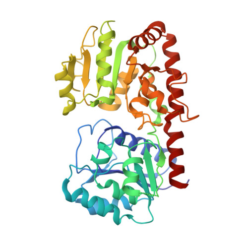
Structural evidence of a passive base-flipping mechanism for AGT, an unusual GT-B glycosyltransferase.

Lariviere, L.Sommer, N.Morera, S.

(2005) J Mol Biol 352: 139-150

  • DOI: https://doi.org/10.1016/j.jmb.2005.07.007
  • Primary Citation of Related Structures:  
    1XV5, 1Y6F, 1Y6G, 1Y8Z, 1YA6

  • PubMed Abstract: 

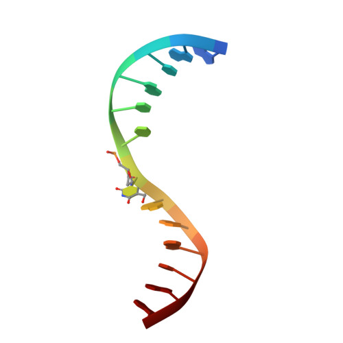
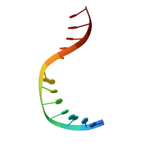
    The Escherichia coli T4 bacteriophage uses two glycosyltransferases to glucosylate and thus protect its DNA: the retaining alpha-glucosyltransferase (AGT) and the inverting beta-glucosyltransferase (BGT). They glucosylate 5-hydroxymethyl cytosine (5-HMC) bases of duplex DNA using UDP-glucose as the sugar donor to form an alpha-glucosidic linkage and a beta-glucosidic linkage, respectively. Five structures of AGT have been determined: a binary complex with the UDP product and four ternary complexes with UDP or UDP-glucose and oligonucleotides containing an A:G, HMU:G (hydroxymethyl uracyl) or AP:G (apurinic/apyrimidinic) mismatch at the target base-pair. AGT adopts the GT-B fold, one of the two folds known for GTs. However, while the sugar donor binding mode is classical for a GT-B enzyme, the sugar acceptor binding mode is unexpected and breaks the established consensus: AGT is the first GT-B enzyme that predominantly binds both the sugar donor and acceptor to the C-terminal domain. Its active site pocket is highly similar to four retaining GT-B glycosyltransferases (trehalose-6-phosphate synthase, glycogen synthase, glycogen and maltodextrin phosphorylases) strongly suggesting a common evolutionary origin and catalytic mechanism for these enzymes. Structure-guided mutagenesis and kinetic analysis do not permit identification of a nucleophile residue responsible for a glycosyl-enzyme intermediate for the classical double displacement mechanism. Interestingly, the DNA structures reveal partially flipped-out bases. They provide evidence for a passive role of AGT in the base-flipping mechanism and for its specific recognition of the acceptor base.


  • Organizational Affiliation

    Laboratoire d'Enzymologie et Biochimie Structurales, UPR 9063 CNRS, Bât.34, 91198-Gif-sur-Yvette, France.


Macromolecules

Find similar proteins by:  (by identity cutoff)  |  3D Structure
Entity ID: 3
MoleculeChains Sequence LengthOrganismDetailsImage
DNA alpha-glucosyltransferaseC [auth A],
D [auth B]
403Tequatrovirus T4Mutation(s): 0 
EC: 2.4.1.26
UniProt
Find proteins for P04519 (Enterobacteria phage T4)
Explore P04519 
Go to UniProtKB:  P04519
Entity Groups  
Sequence Clusters30% Identity50% Identity70% Identity90% Identity95% Identity100% Identity
UniProt GroupP04519
Sequence Annotations
Expand
  • Reference Sequence

Find similar nucleic acids by:  Sequence   |   3D Structure  

Entity ID: 1
MoleculeChains LengthOrganismImage
5'-D(*GP*AP*TP*AP*CP*TP*(5HU)P*AP*GP*AP*TP*AP*G)-3'A [auth C]13N/A
Sequence Annotations
Expand
  • Reference Sequence

Find similar nucleic acids by:  Sequence   |   3D Structure  

Entity ID: 2
MoleculeChains LengthOrganismImage
5'-D(*CP*TP*AP*TP*CP*TP*GP*AP*GP*TP*AP*T)-3'B [auth D]12N/A
Sequence Annotations
Expand
  • Reference Sequence
Small Molecules
Ligands 4 Unique
IDChains Name / Formula / InChI Key2D Diagram3D Interactions
UDP
Query on UDP

Download Ideal Coordinates CCD File 
I [auth A],
N [auth B]
URIDINE-5'-DIPHOSPHATE
C9 H14 N2 O12 P2
XCCTYIAWTASOJW-XVFCMESISA-N
NCO
Query on NCO

Download Ideal Coordinates CCD File 
E [auth D]
F [auth A]
G [auth A]
H [auth A]
K [auth B]
E [auth D],
F [auth A],
G [auth A],
H [auth A],
K [auth B],
L [auth B],
M [auth B]
COBALT HEXAMMINE(III)
Co H18 N6
DYLMFCCYOUSRTK-UHFFFAOYSA-N
EDO
Query on EDO

Download Ideal Coordinates CCD File 
O [auth B]1,2-ETHANEDIOL
C2 H6 O2
LYCAIKOWRPUZTN-UHFFFAOYSA-N
CL
Query on CL

Download Ideal Coordinates CCD File 
J [auth B]CHLORIDE ION
Cl
VEXZGXHMUGYJMC-UHFFFAOYSA-M
Experimental Data & Validation

Experimental Data

  • Method: X-RAY DIFFRACTION
  • Resolution: 2.80 Å
  • R-Value Free: 0.287 
  • R-Value Work: 0.219 
  • Space Group: P 1 21 1
Unit Cell:
Length ( Å )Angle ( ˚ )
a = 46.994α = 90
b = 120.52β = 94.68
c = 86.807γ = 90
Software Package:
Software NamePurpose
MOSFLMdata reduction
SCALAdata scaling
AMoREphasing
CNSrefinement
CCP4data scaling

Structure Validation

View Full Validation Report


Ligand Structure Quality Assessment 


Entry History 

Deposition Data

Revision History  (Full details and data files)

  • Version 1.0: 2005-08-30
    Type: Initial release
  • Version 1.1: 2008-04-30
    Changes: Version format compliance
  • Version 1.2: 2011-07-13
    Changes: Version format compliance
  • Version 1.3: 2020-07-22
    Changes: Advisory, Database references, Derived calculations
  • Version 1.4: 2023-10-25
    Changes: Data collection, Database references, Derived calculations, Refinement description