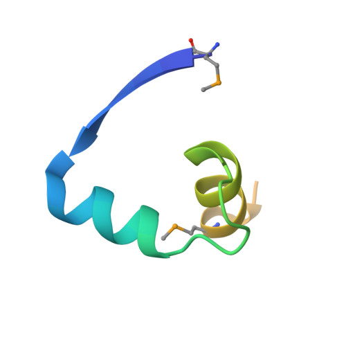
1XRX

Crystal structure of a DNA-binding protein


Experimental Data Snapshot

  • Method: X-RAY DIFFRACTION
  • Resolution: 2.15 Å
  • R-Value Free: 0.237 
  • R-Value Work: 0.222 

wwPDB Validation   3D Report Full Report


This is version 1.3 of the entry. See complete history


Literature

Crystal structure of a SeqA-N filament: implications for DNA replication and chromosome organization.

Guarne, A.Brendler, T.Zhao, Q.Ghirlando, R.Austin, S.Yang, W.

(2005) EMBO J 24: 1502-1511

  • DOI: https://doi.org/10.1038/sj.emboj.7600634
  • Primary Citation of Related Structures:  
    1XRX

  • PubMed Abstract: 

    Escherichia coli SeqA binds clusters of transiently hemimethylated GATC sequences and sequesters the origin of replication, oriC, from methylation and premature reinitiation. Besides oriC, SeqA binds and organizes newly synthesized DNA at replication forks. Binding to multiple GATC sites is crucial for the formation of stable SeqA-DNA complexes. Here we report the crystal structure of the oligomerization domain of SeqA (SeqA-N). The structural unit of SeqA-N is a dimer, which oligomerizes to form a filament. Mutations that disrupt filament formation lead to asynchronous DNA replication, but the resulting SeqA dimer can still bind two GATC sites separated from 5 to 34 base pairs. Truncation of the linker between the oligomerization and DNA-binding domains restricts SeqA to bind two GATC sites separated by one or two full turns. We propose a model of a SeqA filament interacting with multiple GATC sites that accounts for both origin sequestration and chromosome organization.


  • Organizational Affiliation

    Laboratory of Molecular Biology, National Institute of Diabetes, Digestive and Kidney Diseases, National Institutes of Health, Bethesda, MD, USA. guarnea@mcmaster.ca


Macromolecules
Find similar proteins by:  (by identity cutoff)  |  3D Structure
Entity ID: 1
MoleculeChains Sequence LengthOrganismDetailsImage
SeqA protein
A, B, C, D
50Escherichia coliMutation(s): 2 
Gene Names: seqA
UniProt
Find proteins for P0AFY8 (Escherichia coli (strain K12))
Explore P0AFY8 
Go to UniProtKB:  P0AFY8
Entity Groups  
Sequence Clusters30% Identity50% Identity70% Identity90% Identity95% Identity100% Identity
UniProt GroupP0AFY8
Sequence Annotations
Expand
  • Reference Sequence
Small Molecules
Ligands 1 Unique
IDChains Name / Formula / InChI Key2D Diagram3D Interactions
CA
Query on CA

Download Ideal Coordinates CCD File 
E [auth A]CALCIUM ION
Ca
BHPQYMZQTOCNFJ-UHFFFAOYSA-N
Modified Residues  1 Unique
IDChains TypeFormula2D DiagramParent
MSE
Query on MSE
A, B, C, D
L-PEPTIDE LINKINGC5 H11 N O2 SeMET
Experimental Data & Validation

Experimental Data

  • Method: X-RAY DIFFRACTION
  • Resolution: 2.15 Å
  • R-Value Free: 0.237 
  • R-Value Work: 0.222 
  • Space Group: P 31 2 1
Unit Cell:
Length ( Å )Angle ( ˚ )
a = 112.179α = 90
b = 112.179β = 90
c = 62.021γ = 120
Software Package:
Software NamePurpose
CNSrefinement
HKL-2000data reduction
SCALEPACKdata scaling
SOLVEphasing

Structure Validation

View Full Validation Report


Entry History 

Deposition Data

Revision History  (Full details and data files)

  • Version 1.0: 2005-05-10
    Type: Initial release
  • Version 1.1: 2008-04-30
    Changes: Version format compliance
  • Version 1.2: 2011-07-13
    Changes: Derived calculations, Version format compliance
  • Version 1.3: 2013-03-06
    Changes: Other