Single mutation at P1 of a chymotrypsin inhibitor changes it to a trypsin inhibitor: X-ray structural (2.15 A) and biochemical basis
Khamrui, S., Dasgupta, J., Dattagupta, J.K., Sen, U.(2005) Biochim Biophys Acta 1752: 65-72
- PubMed: 16081330
- DOI: https://doi.org/10.1016/j.bbapap.2005.06.012
- Primary Citation of Related Structures:
1XG6 - PubMed Abstract:
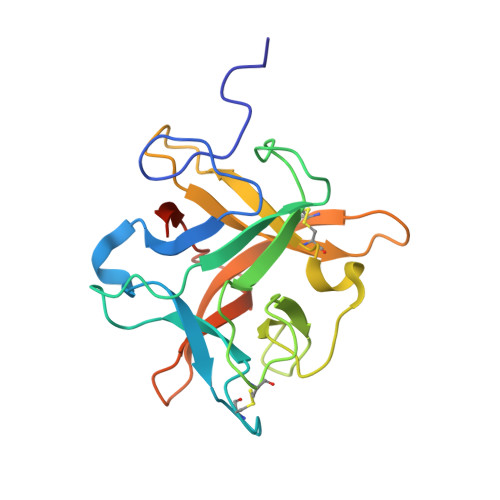
Change in specificity, caused by the mutations at P1 site, of the serine protease inhibitors of different families is reported in the literature, but Kunitz (STI) family inhibitors are almost unexplored in this regard. In this paper, we present the crystal structure of a P1 variant of winged bean chymotrypsin inhibitor (WCI) belonging to Kunitz (STI) family, supplemented by biochemical, phylogenetic and docking studies on the mutant. A single mutation (Leu-->Arg) at P1 converted WCI to a strong inhibitor of trypsin with an association constant of 4.8x10(10) M(-1) which is comparable to other potent trypsin inhibitors of the family. The crystal structure (2.15 A) of this mutant (L65R) shows that its reactive site loop conformation deviates from that of WCI and adopts a structure similar to that of Erythrina caffra trypsin inhibitor (ETI) belonging to the same family. Mutation induced structural changes have also been propagated in a concerted manner to the neighboring conserved scaffolding residue Asn14, such that the side chain of this residue took an orientation similar to that of ETI and optimized the hydrogen bonds with the loop residues. While docking studies provide information about the accommodation of non-specific residues in the active site groove of trypsin, the basis of the directional alteration of the reactive site loop conformation has been understood through sequence analysis and related phylogenetic studies.
Organizational Affiliation:
Crystallography and Molecular Biology Division, Saha Institute of Nuclear Physics, 1/AF Bidhan Nagar, Kolkata 700 064, India.