1XF1

Structure of C5a peptidase- a key virulence factor from Streptococcus


Experimental Data Snapshot

  • Method: X-RAY DIFFRACTION
  • Resolution: 1.90 Å
  • R-Value Free: 0.269 
  • R-Value Work: 0.229 

wwPDB Validation   3D Report Full Report


This is version 1.3 of the entry. See complete history


Literature

Structure of the streptococcal cell wall C5a peptidase

Brown, C.K.Gu, Z.Y.Matsuka, Y.V.Purushothaman, S.S.Winter, L.A.Cleary, P.P.Olmsted, S.B.Ohlendorf, D.H.Earhart, C.A.

(2005) Proc Natl Acad Sci U S A 102: 18391-18396

  • DOI: https://doi.org/10.1073/pnas.0504954102
  • Primary Citation of Related Structures:  
    1XF1

  • PubMed Abstract: 

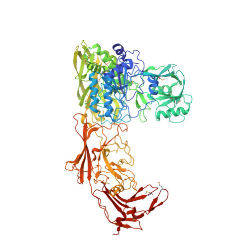
    The structure of a cell surface enzyme from a gram-positive pathogen has been determined to 2-A resolution. Gram-positive pathogens have a thick cell wall to which proteins and carbohydrate are covalently attached. Streptococcal C5a peptidase (SCP), is a highly specific protease and adhesin/invasin. Structural analysis of a 949-residue fragment of the [D130A,S512A] mutant of SCP from group B Streptococcus (S. agalactiae, SCPB) revealed SCPB is composed of five distinct domains. The N-terminal subtilisin-like protease domain has a 134-residue protease-associated domain inserted into a loop between two beta-strands. This domain also contains one of two Arg-Gly-Asp (RGD) sequences found in SCPB. At the C terminus are three fibronectin type III (Fn) domains. The second RGD sequence is located between Fn1 and Fn2. Our analysis suggests that SCP binding to integrins by the RGD motifs may stabilize conformational changes required for substrate binding.


  • Organizational Affiliation

    Department of Biochemistry, Molecular Biology, and Biophysics, University of Minnesota, Minneapolis, MN 55455, USA.


Macromolecules
Find similar proteins by:  (by identity cutoff)  |  3D Structure
Entity ID: 1
MoleculeChains Sequence LengthOrganismDetailsImage
C5a peptidase
A, B
926Streptococcus pyogenesMutation(s): 16 
Gene Names: scpA
EC: 3.4.21
UniProt
Find proteins for P15926 (Streptococcus pyogenes)
Explore P15926 
Go to UniProtKB:  P15926
Entity Groups  
Sequence Clusters30% Identity50% Identity70% Identity90% Identity95% Identity100% Identity
UniProt GroupP15926
Sequence Annotations
Expand
  • Reference Sequence
Small Molecules
Modified Residues  1 Unique
IDChains TypeFormula2D DiagramParent
MSE
Query on MSE
A, B
L-PEPTIDE LINKINGC5 H11 N O2 SeMET
Experimental Data & Validation

Experimental Data

  • Method: X-RAY DIFFRACTION
  • Resolution: 1.90 Å
  • R-Value Free: 0.269 
  • R-Value Work: 0.229 
  • Space Group: P 1 21 1
Unit Cell:
Length ( Å )Angle ( ˚ )
a = 114.696α = 90
b = 75.113β = 104.95
c = 132.391γ = 90
Software Package:
Software NamePurpose
HKL-2000data collection
SCALEPACKdata scaling
SOLVEphasing
CNSrefinement
HKL-2000data reduction

Structure Validation

View Full Validation Report


Entry History 

Deposition Data

Revision History  (Full details and data files)

  • Version 1.0: 2005-11-22
    Type: Initial release
  • Version 1.1: 2008-04-30
    Changes: Version format compliance
  • Version 1.2: 2011-07-13
    Changes: Version format compliance
  • Version 1.3: 2021-10-20
    Changes: Database references, Derived calculations