1XA6

Crystal Structure of the Human Beta2-Chimaerin


Experimental Data Snapshot

  • Method: X-RAY DIFFRACTION
  • Resolution: 3.20 Å
  • R-Value Free: 0.298 
  • R-Value Work: 0.252 
  • R-Value Observed: 0.260 

wwPDB Validation   3D Report Full Report


This is version 1.3 of the entry. See complete history


Literature

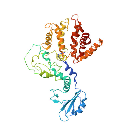
Structural mechanism for lipid activation of the Rac-specific GAP, beta2-chimaerin.

Canagarajah, B.Leskow, F.C.Ho, J.Y.Mischak, H.Saidi, L.F.Kazanietz, M.G.Hurley, J.H.

(2004) Cell 119: 407-418

  • DOI: https://doi.org/10.1016/j.cell.2004.10.012
  • Primary Citation of Related Structures:  
    1XA6

  • PubMed Abstract: 

    The lipid second messenger diacylglycerol acts by binding to the C1 domains of target proteins, which translocate to cell membranes and are allosterically activated. Here we report the crystal structure at 3.2 A resolution of one such protein, beta2-chimaerin, a GTPase-activating protein for the small GTPase Rac, in its inactive conformation. The structure shows that in the inactive state, the N terminus of beta2-chimaerin protrudes into the active site of the RacGAP domain, sterically blocking Rac binding. The diacylglycerol and phospholipid membrane binding site on the C1 domain is buried by contacts with the four different regions of beta2-chimaerin: the N terminus, SH2 domain, RacGAP domain, and the linker between the SH2 and C1 domains. Phospholipid binding to the C1 domain triggers the cooperative dissociation of these interactions, allowing the N terminus to move out of the active site and thereby activating the enzyme.


  • Organizational Affiliation

    Laboratory of Molecular Biology, National Institute of Diabetes and Digestive and Kidney Diseases, National Institutes of Health, US Department of Health and Human Services, Bethesda, MD 20892, USA.


Macromolecules
Find similar proteins by:  (by identity cutoff)  |  3D Structure
Entity ID: 1
MoleculeChains Sequence LengthOrganismDetailsImage
Beta2-chimaerin466Homo sapiensMutation(s): 0 
Membrane Entity: Yes 
UniProt & NIH Common Fund Data Resources
Find proteins for P52757 (Homo sapiens)
Explore P52757 
Go to UniProtKB:  P52757
PHAROS:  P52757
GTEx:  ENSG00000106069 
Entity Groups  
Sequence Clusters30% Identity50% Identity70% Identity90% Identity95% Identity100% Identity
UniProt GroupP52757
Sequence Annotations
Expand
  • Reference Sequence
Experimental Data & Validation

Experimental Data

  • Method: X-RAY DIFFRACTION
  • Resolution: 3.20 Å
  • R-Value Free: 0.298 
  • R-Value Work: 0.252 
  • R-Value Observed: 0.260 
  • Space Group: P 61 2 2
Unit Cell:
Length ( Å )Angle ( ˚ )
a = 131.09α = 90
b = 131.09β = 90
c = 288.75γ = 120
Software Package:
Software NamePurpose
DENZOdata reduction
SCALEPACKdata scaling
SOLVEphasing
CNSrefinement

Structure Validation

View Full Validation Report


Entry History 

Deposition Data

Revision History  (Full details and data files)

  • Version 1.0: 2004-11-23
    Type: Initial release
  • Version 1.1: 2008-04-30
    Changes: Version format compliance
  • Version 1.2: 2011-07-13
    Changes: Version format compliance
  • Version 1.3: 2024-02-14
    Changes: Data collection, Database references, Derived calculations