1X2E

The crystal structure of prolyl aminopeptidase complexed with Ala-TBODA


Experimental Data Snapshot

  • Method: X-RAY DIFFRACTION
  • Resolution: 2.40 Å
  • R-Value Free: 0.217 
  • R-Value Work: 0.177 

wwPDB Validation   3D Report Full Report


Ligand Structure Quality Assessment 


This is version 1.3 of the entry. See complete history


Literature

Unusual extra space at the active site and high activity for acetylated hydroxyproline of prolyl aminopeptidase from Serratia marcescens

Nakajima, Y.Ito, K.Sakata, M.Xu, Y.Nakashima, K.Matsubara, F.Hatakeyama, S.Yoshimoto, T.

(2006) J Bacteriol 188: 1599-1606

  • DOI: https://doi.org/10.1128/JB.188.4.1599-1606.2006
  • Primary Citation of Related Structures:  
    1X2B, 1X2E

  • PubMed Abstract: 

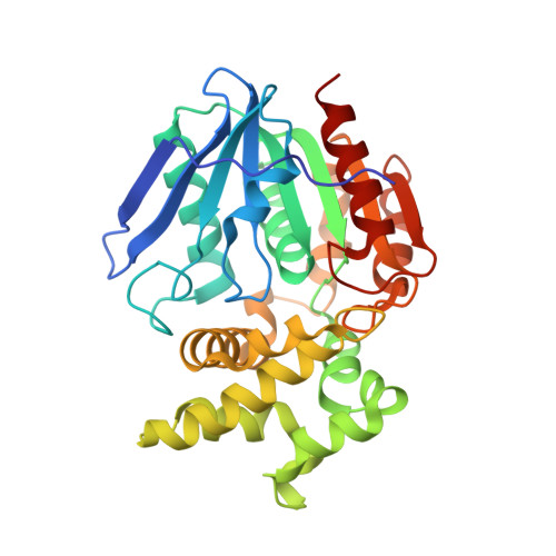
    The prolyl aminopeptidase complexes of Ala-TBODA [2-alanyl-5-tert-butyl-(1, 3, 4)-oxadiazole] and Sar-TBODA [2-sarcosyl-5-tert-butyl-(1, 3, 4)-oxadiazole] were analyzed by X-ray crystallography at 2.4 angstroms resolution. Frames of alanine and sarcosine residues were well superimposed on each other in the pyrrolidine ring of proline residue, suggesting that Ala and Sar are recognized as parts of this ring of proline residue by the presence of a hydrophobic proline pocket at the active site. Interestingly, there was an unusual extra space at the bottom of the hydrophobic pocket where proline residue is fixed in the prolyl aminopeptidase. Moreover, 4-acetyloxyproline-betaNA (4-acetyloxyproline beta-naphthylamide) was a better substrate than Pro-betaNA. Computer docking simulation well supports the idea that the 4-acetyloxyl group of the substrate fitted into that space. Alanine scanning mutagenesis of Phe139, Tyr149, Tyr150, Phe236, and Cys271, consisting of the hydrophobic pocket, revealed that all of these five residues are involved significantly in the formation of the hydrophobic proline pocket for the substrate. Tyr149 and Cys271 may be important for the extra space and may orient the acetyl derivative of hydroxyproline to a preferable position for hydrolysis. These findings imply that the efficient degradation of collagen fragment may be achieved through an acetylation process by the bacteria.


  • Organizational Affiliation

    Graduate School of Biomedical Sciences, Nagasaki University, Nagasaki 852-8521, Japan.


Macromolecules
Find similar proteins by:  (by identity cutoff)  |  3D Structure
Entity ID: 1
MoleculeChains Sequence LengthOrganismDetailsImage
Proline iminopeptidase317Serratia marcescensMutation(s): 0 
EC: 3.4.11.5
UniProt
Find proteins for O32449 (Serratia marcescens)
Explore O32449 
Go to UniProtKB:  O32449
Entity Groups  
Sequence Clusters30% Identity50% Identity70% Identity90% Identity95% Identity100% Identity
UniProt GroupO32449
Sequence Annotations
Expand
  • Reference Sequence
Small Molecules
Ligands 1 Unique
IDChains Name / Formula / InChI Key2D Diagram3D Interactions
ATX
Query on ATX

Download Ideal Coordinates CCD File 
B [auth A](2S)-2-AMINO-1-(5-TERT-BUTYL-1,3,4-OXADIAZOL-2-YL)PROPAN-1-ONE
C9 H15 N3 O2
PVDZDTVFUVTTDU-YFKPBYRVSA-N
Experimental Data & Validation

Experimental Data

  • Method: X-RAY DIFFRACTION
  • Resolution: 2.40 Å
  • R-Value Free: 0.217 
  • R-Value Work: 0.177 
  • Space Group: P 43 21 2
Unit Cell:
Length ( Å )Angle ( ˚ )
a = 65.187α = 90
b = 65.187β = 90
c = 169.282γ = 90
Software Package:
Software NamePurpose
HKL-2000data collection
SCALEPACKdata scaling
CNSrefinement
HKL-2000data reduction
CNSphasing

Structure Validation

View Full Validation Report


Ligand Structure Quality Assessment 


Entry History 

Deposition Data

Revision History  (Full details and data files)

  • Version 1.0: 2006-05-09
    Type: Initial release
  • Version 1.1: 2008-04-30
    Changes: Version format compliance
  • Version 1.2: 2011-07-13
    Changes: Version format compliance
  • Version 1.3: 2023-10-25
    Changes: Data collection, Database references, Derived calculations, Refinement description