Comparison of crystal structures of two homologous proteins: structural origin of altered domain interactions in immunoglobulin light-chain dimers.
Huang, D.B., Chang, C.H., Ainsworth, C., Brunger, A.T., Eulitz, M., Solomon, A., Stevens, F.J., Schiffer, M.(1994) Biochemistry 33: 14848-14857
- PubMed: 7993911
- DOI: https://doi.org/10.1021/bi00253a024
- Primary Citation of Related Structures:
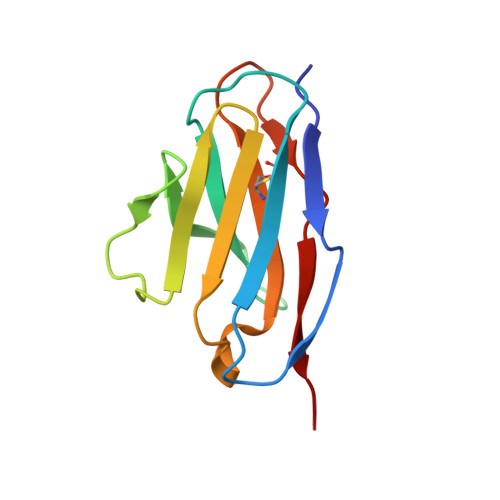
1WTL - PubMed Abstract:
The sequence and structure of a second human kappa 1 immunoglobulin light-chain variable domain, Wat, has been determined. The R-factor is 15.7% for 1.9-A data. One hundred and ninety-five water molecules were identified; 30 water molecules were located in identical positions in each of the monomers. Some of the water molecules are integral parts of the domains. This light chain is encoded by the same variable domain gene that encoded the previously characterized kappa I variable domain, Rei. Due to limited somatic mutation, the two highly homologous proteins differ in only 20 of the 108 residues. Wat crystallized in space group P6(4) while Rei crystallized in space group P6(1); in both crystals, the asymmetric unit was the noncovalent dimer. Although the basic domain structure is the same for both proteins, the relative positions of the domains within the two dimers differ. This difference is most likely accounted for by the replacement of Tyr36 in Rei by Phe in the Wat protein. Residue Tyr36 is part of the hydrogen-bonding network in the interface between the domains in Rei. Losing the hydrogen-bonding capability of residue 36 by replacement of Tyr by Phe alters the network of hydrogen bonds between the domains, resulting in a different domain-domain contact. The details of lattice contacts in the two crystals were compared. One type of contact that extends the beta-sheet of the individual domains was conserved, but because it involved different symmetry elements within the crystal, different crystal packing resulted. In the Wat crystal, one of the contacts shows an example of how a symmetrical binding site can "bind" an asymmetrical object. Further, the examination of the Wat crystal also illustrates how the different crystalline environments of the domains of the dimer results in different distributions of temperature factors for the residues within the domains.
Organizational Affiliation:
Center for Mechanistic Biology and Biotechnology, Argonne National Laboratory, Illinois 60439-4833.