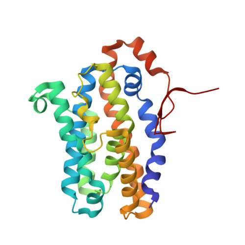
Crystal structure of dimeric heme oxygenase-2 from Synechocystis sp. PCC 6803 in complex with heme.
Sugishima, M., Hagiwara, Y., Zhang, X., Yoshida, T., Migita, C.T., Fukuyama, K.(2005) Biochemistry 44: 4257-4266
- PubMed: 15766254
- DOI: https://doi.org/10.1021/bi0480483
- Primary Citation of Related Structures:
1WOV, 1WOW, 1WOX - PubMed Abstract:
Phycobiliproteins, light-harvesting proteins in cyanobacteria, red algae, and cryptophytes, contain phycobilin pigments. Phycobilins are synthesized from biliverdin, which is produced by the oxidative cleavage of the heme porphyrin ring catalyzed by heme oxygenase (HO). Two paralogs of ho (ho1 and ho2) have been identified in the genome of the cyanobacterium, Synechocystis sp. PCC 6803. The recombinant proteins of both paralogs (Syn HO-1 and Syn HO-2) possess in vitro heme degradation activity. We have determined the crystal structures of Syn HO-2 in complex with heme (heme-Syn HO-2) and its reduced and NO bound forms. The heme-Syn HO-2 crystal was a nonmerohedral twin, and detwinned diffraction data were used to refine the structure. Although heme-Syn HO-2 shares common folding with other HOs, the C-terminal segment is ordered and turns back to the heme-binding side. Gel-filtration chromatography analysis and molecular packing in the crystal indicate that heme-Syn HO-2 forms a homodimer, in which the C-terminal ordered segments interact with each other. Because Syn HO-2 is a monomer in the apo state, the dimeric interaction may aid in the selection of the reducing partner but likely does not interfere with heme binding. The heme iron is coordinated by a water molecule in the ferric form, but the distal water is absent in the ferrous form. In all of the Syn HO-2 structures, several water molecules form a hydrogen-bond network at the distal hemepocket, which is involved in HO activity. Upon NO binding, the side-chain conformation of Tyr 156 changes. Tyr 156 is located at the hydrophobic cluster, which interrupts the possible H(+) pathway from the molecular surface to the hemepocket. Thus, Tyr 156 may function as a H(+) shuttle by changing conformation.
Organizational Affiliation:
Department of Biology, Graduate School of Science, Osaka University, Toyonaka, Osaka 560-0043, Japan.