1W9T

Structure of a beta-1,3-glucan binding CBM6 from Bacillus halodurans in complex with xylobiose


Experimental Data Snapshot

  • Method: X-RAY DIFFRACTION
  • Resolution: 1.62 Å
  • R-Value Free: 0.179 
  • R-Value Work: 0.118 
  • R-Value Observed: 0.121 

wwPDB Validation   3D Report Full Report


Ligand Structure Quality Assessment 


This is version 2.1 of the entry. See complete history


Literature

Family 6 Carbohydrate Binding Modules Recognize the Non-Reducing End of Beta-1,3-Linked Glucans by Presenting a Unique Ligand Binding Surface

Van Bueren, A.L.Moreland, C.Gilbert, H.J.Boraston, A.B.

(2005) J Biol Chem 280: 530

  • DOI: https://doi.org/10.1074/jbc.M410113200
  • Primary Citation of Related Structures:  
    1W9S, 1W9T, 1W9W

  • PubMed Abstract: 

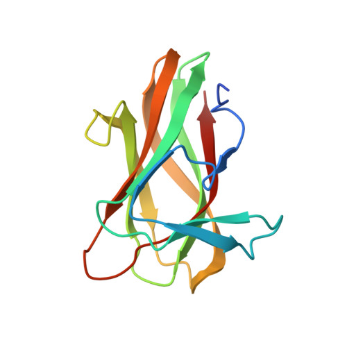
    Enzymes that hydrolyze insoluble complex polysaccharide structures contain non-catalytic carbohydrate binding modules (CBMS) that play a pivotal role in the action of these enzymes against recalcitrant substrates. Family 6 CBMs (CBM6s) are distinct from other CBM families in that these protein modules contain multiple distinct ligand binding sites, a feature that makes CBM6s particularly appropriate receptors for the beta-1,3-glucan laminarin, which displays an extended U-shaped conformation. To investigate the mechanism by which family 6 CBMs recognize laminarin, we report the biochemical and structural properties of a CBM6 (designated BhCBM6) that is located in an enzyme, which is shown, in this work, to display beta-1,3-glucanase activity. BhCBM6 binds beta-1,3-glucooligosaccharides with affinities of approximately 1 x 10(5) m(-1). The x-ray crystal structure of this CBM in complex with laminarihexaose reveals similarity with the structures of other CBM6s but a unique binding mode. The binding cleft in this protein is sealed at one end, which prevents binding of linear polysaccharides such as cellulose, and the orientation of the sugar at this site prevents glycone extension of the ligand and thus conferring specificity for the non-reducing ends of glycans. The high affinity for extended beta-1,3-glucooligosaccharides is conferred by interactions with the surface of the protein located between the two binding sites common to CBM6s and thus reveals a third ligand binding site in family 6 CBMs. This study therefore demonstrates how the multiple binding clefts and highly unusual protein surface of family 6 CBMs confers the extensive range of specificities displayed by this protein family. This is in sharp contrast to other families of CBMs where variation in specificity between different members reflects differences in the topology of a single binding site.


  • Organizational Affiliation

    Department of Biochemistry and Microbiology, University of Victoria, British Columbia V8W 3P6, Canada.


Macromolecules
Find similar proteins by:  (by identity cutoff)  |  3D Structure
Entity ID: 1
MoleculeChains Sequence LengthOrganismDetailsImage
BH0236 PROTEIN
A, B
142Halalkalibacterium haloduransMutation(s): 0 
UniProt
Find proteins for Q9KG76 (Halalkalibacterium halodurans (strain ATCC BAA-125 / DSM 18197 / FERM 7344 / JCM 9153 / C-125))
Explore Q9KG76 
Go to UniProtKB:  Q9KG76
Entity Groups  
Sequence Clusters30% Identity50% Identity70% Identity90% Identity95% Identity100% Identity
UniProt GroupQ9KG76
Sequence Annotations
Expand
  • Reference Sequence
Oligosaccharides

Help

Entity ID: 2
MoleculeChains Length2D Diagram Glycosylation3D Interactions
beta-D-xylopyranose-(1-4)-beta-D-xylopyranose
C, D
2N/A
Glycosylation Resources
GlyTouCan:  G87728WL
GlyCosmos:  G87728WL
Small Molecules
Ligands 3 Unique
IDChains Name / Formula / InChI Key2D Diagram3D Interactions
XYS
Query on XYS

Download Ideal Coordinates CCD File 
H [auth A],
N [auth B]
alpha-D-xylopyranose
C5 H10 O5
SRBFZHDQGSBBOR-LECHCGJUSA-N
XYP
Query on XYP

Download Ideal Coordinates CCD File 
I [auth A],
J [auth A],
O [auth B],
P [auth B]
beta-D-xylopyranose
C5 H10 O5
SRBFZHDQGSBBOR-KKQCNMDGSA-N
NA
Query on NA

Download Ideal Coordinates CCD File 
E [auth A]
F [auth A]
G [auth A]
K [auth B]
L [auth B]
E [auth A],
F [auth A],
G [auth A],
K [auth B],
L [auth B],
M [auth B]
SODIUM ION
Na
FKNQFGJONOIPTF-UHFFFAOYSA-N
Biologically Interesting Molecules (External Reference) 1 Unique
Experimental Data & Validation

Experimental Data

  • Method: X-RAY DIFFRACTION
  • Resolution: 1.62 Å
  • R-Value Free: 0.179 
  • R-Value Work: 0.118 
  • R-Value Observed: 0.121 
  • Space Group: P 1
Unit Cell:
Length ( Å )Angle ( ˚ )
a = 30.711α = 108.89
b = 40.967β = 105.87
c = 56.039γ = 90.03
Software Package:
Software NamePurpose
REFMACrefinement
MOLREPphasing

Structure Validation

View Full Validation Report


Ligand Structure Quality Assessment 


Entry History 

Deposition Data

Revision History  (Full details and data files)

  • Version 1.0: 2004-11-03
    Type: Initial release
  • Version 1.1: 2013-08-07
    Changes: Derived calculations, Other, Refinement description, Structure summary, Version format compliance
  • Version 2.0: 2020-07-29
    Type: Remediation
    Reason: Carbohydrate remediation
    Changes: Advisory, Atomic model, Data collection, Derived calculations, Other, Structure summary
  • Version 2.1: 2023-12-13
    Changes: Data collection, Database references, Derived calculations, Refinement description, Structure summary