1W9A

Crystal structure of Rv1155 from Mycobacterium tuberculosis


Experimental Data Snapshot

  • Method: X-RAY DIFFRACTION
  • Resolution: 1.80 Å
  • R-Value Free: 0.175 
  • R-Value Work: 0.141 
  • R-Value Observed: 0.143 

wwPDB Validation   3D Report Full Report


This is version 1.2 of the entry. See complete history


Literature

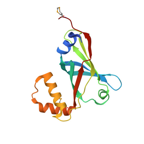
Crystal Structure of the Conserved Hypothetical Protein Rv1155 from Mycobacterium Tuberculosis

Canaan, S.Sulzenbacher, G.Roig-Zamboni, V.Scappuccini-Calvo, L.Frassinetti, F.Maurin, D.Cambillau, C.Bourne, Y.

(2005) FEBS Lett 579: 215

  • DOI: https://doi.org/10.1016/j.febslet.2004.11.069
  • Primary Citation of Related Structures:  
    1W9A

  • PubMed Abstract: 

    With the aim of elucidating the biological function of hypothetical proteins unique amongst the Actynomyces sub-group of bacteria, we have solved the crystal structure of the conserved hypothetical protein Rv1155 from Mycobacterium tuberculosis at 1.8 A resolution. Rv1155 is a homodimer both in the crystal structure and in solution and folds into two separate domains consisting of a six-stranded anti-parallel beta-barrel fold flanked by two alpha-helices and a helix-turn-helix domain. Both domains contribute to the formation of two deep clefts at the dimer interface. The overall fold of Rv1155 strikingly resembles that of flavin mononucleotide-binding protein and pyridoxamine 5'-phosphate oxydase, but the architecture of the putative binding pocket is markedly different, consistent with the lack of color of Rv1155 and its inability to bind FMN. Rv1155 thus appears to belong to a group of proteins with stringent conservation of the binding cleft, having evolved towards a new binding function.


  • Organizational Affiliation

    Architecture et Fonction des Macromolécules Biologiques, CNRS UMR-6098, 31 Chemin Joseph Aiguier, F-13402 Marseille Cedex 20, France.


Macromolecules
Find similar proteins by:  (by identity cutoff)  |  3D Structure
Entity ID: 1
MoleculeChains Sequence LengthOrganismDetailsImage
PUTATIVE PYRIDOXINE/PYRIDOXAMINE 5'-PHOSPHATE OXIDASE
A, B
147Mycobacterium tuberculosis H37RvMutation(s): 0 
EC: 1.4.3.5
UniProt
Find proteins for O06553 (Mycobacterium tuberculosis (strain ATCC 25618 / H37Rv))
Explore O06553 
Go to UniProtKB:  O06553
Entity Groups  
Sequence Clusters30% Identity50% Identity70% Identity90% Identity95% Identity100% Identity
UniProt GroupO06553
Sequence Annotations
Expand
  • Reference Sequence
Small Molecules
Modified Residues  1 Unique
IDChains TypeFormula2D DiagramParent
MSE
Query on MSE
A, B
L-PEPTIDE LINKINGC5 H11 N O2 SeMET
Experimental Data & Validation

Experimental Data

  • Method: X-RAY DIFFRACTION
  • Resolution: 1.80 Å
  • R-Value Free: 0.175 
  • R-Value Work: 0.141 
  • R-Value Observed: 0.143 
  • Space Group: P 1 21 1
Unit Cell:
Length ( Å )Angle ( ˚ )
a = 46.953α = 90
b = 55.122β = 108.31
c = 55.231γ = 90
Software Package:
Software NamePurpose
REFMACrefinement
DENZOdata reduction
SCALAdata scaling
SOLVEphasing

Structure Validation

View Full Validation Report


Entry History 

Deposition Data

Revision History  (Full details and data files)

  • Version 1.0: 2005-01-06
    Type: Initial release
  • Version 1.1: 2011-07-13
    Changes: Advisory, Version format compliance
  • Version 1.2: 2013-03-06
    Changes: Data collection, Other, Source and taxonomy, Structure summary