1W5U

GRAMICIDIN D FROM BACILLUS BREVIS (ETHANOL SOLVATE)


Experimental Data Snapshot

  • Method: X-RAY DIFFRACTION
  • Resolution: 1.14 Å
  • R-Value Free: 0.215 
  • R-Value Observed: 0.171 

wwPDB Validation   3D Report Full Report


This is version 2.1 of the entry. See complete history


Literature

Structure of Gramicidin D-Rbcl Complex at Atomic Resolution from Low-Temperature Synchrotron Data: Interactions of Double-Stranded Gramicidin Channel Contents and Cations with Channel Wall

Glowka, M.L.Olczak, A.Bojarska, J.Szczesio, M.Duax, W.L.Burkhart, B.M.Pangborn, W.A.Langs, D.A.Wawrzak, Z.

(2005) Acta Crystallogr D Biol Crystallogr 61: 433

  • DOI: https://doi.org/10.1107/S0907444905000399
  • Primary Citation of Related Structures:  
    1W5U

  • PubMed Abstract: 

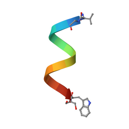
    Gramicidin D (gD) is a naturally occurring ionophoric antibiotic that forms membrane channels specific for monovalent cations. The crystal structure of the RbCl complex of gD has been determined at 1.14 A resolution from low-temperature (100 K) synchrotron-radiation data with a final R of 16%. The structure was refined with anisotropic temperature factors for all non-H atoms and with partial occupancies for many of them. The asymmetric unit in the crystal contains four crystallographically independent molecules that form two right-handed antiparallel double-stranded dimers. There are seven distinct rubidium-binding sites in each dimeric channel. The occupancy factors of Rb cations are between 0.11 and 0.35 and the total ion contents of the two crystallographically independent channels are 1.59 and 1.22 ions, respectively. Although each channel is 'chemically symmetrical', the side-chain conformations, the distributions of rubidium cations and their binding sites in the two independent channels are not. Cations are 'coordinated' by delocalized pi-electrons of three to five carbonyl groups that together with peptide backbone chains form the gramicidin channel walls. The water:cation ratio in the channel interior is four or five:one, and five or six waters separate Rb cations during their passage through the channel.


  • Organizational Affiliation

    Institute of General and Ecological Chemistry, Technical University, Łódź, Poland. marekglo@p.lodz.pl


Macromolecules

Find similar proteins by:  Sequence   |   3D Structure  

Entity ID: 1
MoleculeChains Sequence LengthOrganismDetailsImage
GRAMICIDIN D
A, B, C, D
16Brevibacillus brevisMutation(s): 0 
Entity Groups  
Sequence Clusters30% Identity50% Identity70% Identity90% Identity95% Identity100% Identity
Sequence Annotations
Expand
  • Reference Sequence
Small Molecules
Ligands 3 Unique
IDChains Name / Formula / InChI Key2D Diagram3D Interactions
RB
Query on RB

Download Ideal Coordinates CCD File 
E [auth A]
F [auth A]
I [auth B]
J [auth B]
K [auth B]
E [auth A],
F [auth A],
I [auth B],
J [auth B],
K [auth B],
O [auth C],
Q [auth C],
R [auth C],
S [auth C],
T [auth C],
U [auth C],
V [auth D],
W [auth D],
X [auth D]
RUBIDIUM ION
Rb
NCCSSGKUIKYAJD-UHFFFAOYSA-N
EOH
Query on EOH

Download Ideal Coordinates CCD File 
P [auth C]ETHANOL
C2 H6 O
LFQSCWFLJHTTHZ-UHFFFAOYSA-N
CL
Query on CL

Download Ideal Coordinates CCD File 
G [auth A]
H [auth A]
L [auth B]
M [auth B]
N [auth B]
G [auth A],
H [auth A],
L [auth B],
M [auth B],
N [auth B],
Y [auth D]
CHLORIDE ION
Cl
VEXZGXHMUGYJMC-UHFFFAOYSA-M
Modified Residues  1 Unique
IDChains TypeFormula2D DiagramParent
FVA
Query on FVA
A, B, C, D
L-PEPTIDE LINKINGC6 H11 N O3VAL
Biologically Interesting Molecules (External Reference) 1 Unique
Experimental Data & Validation

Experimental Data

  • Method: X-RAY DIFFRACTION
  • Resolution: 1.14 Å
  • R-Value Free: 0.215 
  • R-Value Observed: 0.171 
  • Space Group: P 21 21 21
Unit Cell:
Length ( Å )Angle ( ˚ )
a = 30.06α = 90
b = 31.3β = 90
c = 51.691γ = 90
Software Package:
Software NamePurpose
SHELXL-97refinement
DENZOdata reduction
SCALEPACKdata scaling

Structure Validation

View Full Validation Report


Entry History 

Deposition Data

Revision History  (Full details and data files)

  • Version 1.0: 2005-09-15
    Type: Initial release
  • Version 1.1: 2011-07-13
    Changes: Atomic model, Database references, Derived calculations, Structure summary, Version format compliance
  • Version 1.2: 2012-11-30
    Changes: Other
  • Version 1.3: 2019-05-22
    Changes: Data collection, Other, Refinement description
  • Version 2.0: 2023-11-15
    Changes: Atomic model, Data collection, Database references, Derived calculations, Other
  • Version 2.1: 2023-12-13
    Changes: Refinement description