1VPW

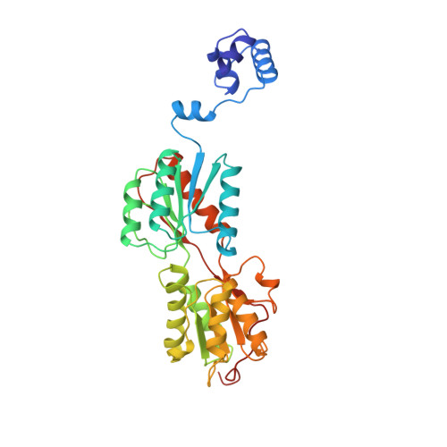
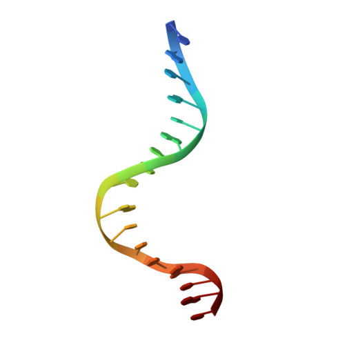
STRUCTURE OF THE PURR MUTANT, L54M, BOUND TO HYPOXANTHINE AND PURF OPERATOR DNA


Experimental Data Snapshot

  • Method: X-RAY DIFFRACTION
  • Resolution: 2.70 Å
  • R-Value Work: 0.174 

wwPDB Validation   3D Report Full Report


This is version 1.5 of the entry. See complete history


Literature

The structure of PurR mutant L54M shows an alternative route to DNA kinking.

Arvidson, D.N.Lu, F.Faber, C.Zalkin, H.Brennan, R.G.

(1998) Nat Struct Biol 5: 436-441

  • DOI: https://doi.org/10.1038/nsb0698-436
  • Primary Citation of Related Structures:  
    1VPW

  • PubMed Abstract: 

    The crystal structure of the purine repressor mutant L54M bound to hypoxanthine and to the purF operator provides a stereochemical understanding of the high DNA affinity of this hinge helix mutant. Comparison of the PurR L54M-DNA complex to that of the wild type PurR-DNA complex reveals that these purine repressors bind and kink DNA similarly despite significant differences in their minor groove contacts and routes to interdigitation of the central C.G:G.C base pair step. Modeling studies, supported by genetic and biochemical data, show that the stereochemistry of the backbone atoms of the abutting hinge helices combined with the rigidity of the kinked base pair step constrain the interdigitating residue to leucine or methionine for the LacI/GalR family of transcription regulators.


  • Organizational Affiliation

    Department of Biochemistry and Molecular Biology, Oregon Health Sciences University, Portland 97201-3098, USA.


Macromolecules

Find similar proteins by:  (by identity cutoff)  |  3D Structure
Entity ID: 2
MoleculeChains Sequence LengthOrganismDetailsImage
PURINE REPRESSORB [auth A]340Escherichia coliMutation(s): 1 
UniProt
Find proteins for P0ACP7 (Escherichia coli (strain K12))
Explore P0ACP7 
Go to UniProtKB:  P0ACP7
Entity Groups  
Sequence Clusters30% Identity50% Identity70% Identity90% Identity95% Identity100% Identity
UniProt GroupP0ACP7
Sequence Annotations
Expand
  • Reference Sequence

Find similar nucleic acids by:  Sequence   |   3D Structure  

Entity ID: 1
MoleculeChains LengthOrganismImage
DNA (5'-D(*TP*AP*CP*GP*CP*AP*AP*AP*CP*GP*TP*TP*TP*GP*CP*GP*T )-3')A [auth B]17synthetic construct
Sequence Annotations
Expand
  • Reference Sequence
Small Molecules
Ligands 1 Unique
IDChains Name / Formula / InChI Key2D Diagram3D Interactions
HPA
Query on HPA

Download Ideal Coordinates CCD File 
C [auth A]HYPOXANTHINE
C5 H4 N4 O
FDGQSTZJBFJUBT-UHFFFAOYSA-N
Experimental Data & Validation

Experimental Data

  • Method: X-RAY DIFFRACTION
  • Resolution: 2.70 Å
  • R-Value Work: 0.174 
  • Space Group: C 2 2 21
Unit Cell:
Length ( Å )Angle ( ˚ )
a = 175.89α = 90
b = 94.93β = 90
c = 81.9γ = 90
Software Package:
Software NamePurpose
TNTrefinement

Structure Validation

View Full Validation Report


Entry History 

Deposition Data

Revision History  (Full details and data files)

  • Version 1.0: 1998-06-17
    Type: Initial release
  • Version 1.1: 2008-05-22
    Changes: Version format compliance
  • Version 1.2: 2011-07-13
    Changes: Version format compliance
  • Version 1.3: 2017-02-15
    Changes: Source and taxonomy, Structure summary
  • Version 1.4: 2021-11-03
    Changes: Database references, Derived calculations
  • Version 1.5: 2024-02-14
    Changes: Data collection