Structural evidence for evolution of shark Ig new antigen receptor variable domain antibodies from a cell-surface receptor
Streltsov, V.A., Varghese, J.N., Carmichael, J.A., Irving, R.A., Hudson, P.J., Nuttall, S.D.(2004) Proc Natl Acad Sci U S A 101: 12444-12449
- PubMed: 15304650
- DOI: https://doi.org/10.1073/pnas.0403509101
- Primary Citation of Related Structures:
1VER, 1VES - PubMed Abstract:
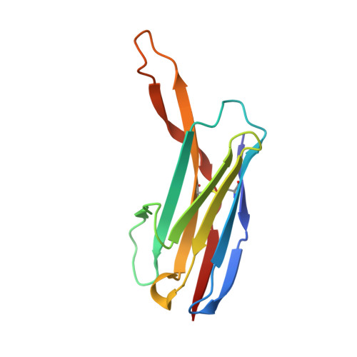
The Ig new antigen receptors (IgNARs) are single-domain antibodies found in the serum of sharks. Here, we report 2.2- and 2.8-A structures of the type 2 IgNAR variable domains 12Y-1 and 12Y-2. Structural features include, first, an Ig superfamily topology transitional between cell adhesion molecules, antibodies, and T cell receptors; and, second, a vestigial complementarity-determining region 2 at the "bottom" of the molecule, apparently discontinuous from the antigen-binding paratope and similar to that observed in cell adhesion molecules. Thus, we suggest that IgNARs originated as cell-surface adhesion molecules coopted to the immune repertoire and represent an evolutionary lineage independent of variable heavy chain/variable light chain type antibodies. Additionally, both 12Y-1 and 12Y-2 form unique crystallographic dimers, predominantly mediated by main-chain framework interactions, which represent a possible model for primordial cell-based interactions. Unusually, the 12Y-2 complementarity-determining region 3 also adopts an extended beta-hairpin structure, suggesting a distinct selective advantage in accessing cryptic antigenic epitopes.
Organizational Affiliation:
Division of Health Sciences and Nutrition, Commonwealth Scientific and Industrial Research Organization, and Cooperative Research Centre for Diagnostics, 343 Royal Parade, Parkville 3052, Australia.