1VEO

Crystal Structure Analysis of Y164F/maltose of Bacillus cereus Beta-Amylase at pH 4.6


Experimental Data Snapshot

  • Method: X-RAY DIFFRACTION
  • Resolution: 2.12 Å
  • R-Value Free: 0.215 
  • R-Value Work: 0.180 
  • R-Value Observed: 0.180 

wwPDB Validation   3D Report Full Report


Ligand Structure Quality Assessment 


This is version 2.2 of the entry. See complete history


Literature

Engineering of the pH optimum of Bacillus cereus beta-amylase: conversion of the pH optimum from a bacterial type to a higher-plant type

Hirata, A.Adachi, M.Utsumi, S.Mikami, B.

(2004) Biochemistry 43: 12523-12531

  • DOI: https://doi.org/10.1021/bi049173h
  • Primary Citation of Related Structures:  
    1VEM, 1VEN, 1VEO, 1VEP

  • PubMed Abstract: 

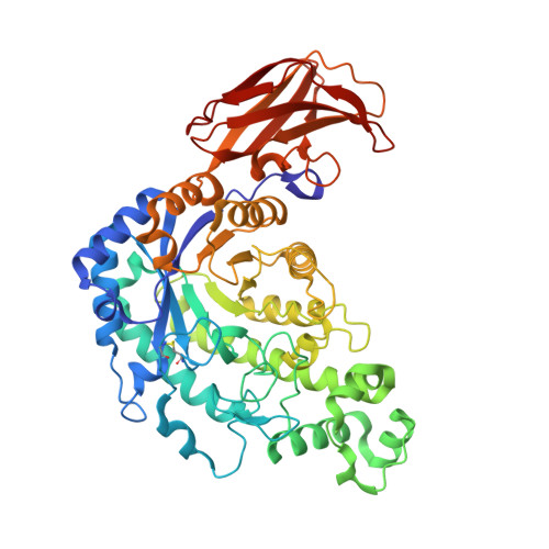
    The optimum pH of Bacillus cereus beta-amylase (BCB, pH 6.7) differs from that of soybean beta-amylase (SBA, pH 5.4) due to the substitution of a few amino acid residues near the catalytic base residue (Glu 380 in SBA and Glu 367 in BCB). To explore the mechanism for controlling the optimum pH of beta-amylase, five mutants of BCB (Y164E, Y164F, Y164H, Y164Q, and Y164Q/T47M/Y164E/T328N) were constructed and characterized with respect to enzymatic properties and X-ray structural crystal analysis. The optimum pH of the four single mutants shifted to 4.2-4.8, approximately 2 pH units and approximately 1 pH unit lower than those of BCB and SBA, respectively, and their k(cat) values decreased to 41-3% of that of the wild-type enzyme. The X-ray crystal analysis of the enzyme-maltose complexes showed that Glu 367 of the wild type is surrounded by two water molecules (W1 and W2) that are not found in SBA. W1 is hydrogen-bonded to both side chains of Glu 367 and Tyr 164. The mutation of Tyr 164 to Glu and Phe resulted in the disruption of the hydrogen bond between Tyr 164 Oeta and W1 and the introduction of two additional water molecules near position 164. In contrast, the triple mutant of BCB with a slightly decreased pH optimum at pH 6.0 has no water molecules (W1 and W2) around Glu 367. These results suggested that a water-mediated hydrogen bond network (Glu 367...W1...Tyr 164...Thr 328) is the primary requisite for the increased pH optimum of wild-type BCB. This strategy is completely different from that of SBA, in which a hydrogen bond network (Glu 380...Thr 340...Glu 178) reduces the optimum pH in a hydrophobic environment.


  • Organizational Affiliation

    Laboratory of Food Quality Design and Development, Graduate School of Agriculture, Kyoto University, Gokasho, Uji, Kyoto 611-0011, Japan.


Macromolecules
Find similar proteins by:  (by identity cutoff)  |  3D Structure
Entity ID: 1
MoleculeChains Sequence LengthOrganismDetailsImage
Beta-amylase516Bacillus cereusMutation(s): 1 
EC: 3.2.1.2
UniProt
Find proteins for P36924 (Bacillus cereus)
Explore P36924 
Go to UniProtKB:  P36924
Entity Groups  
Sequence Clusters30% Identity50% Identity70% Identity90% Identity95% Identity100% Identity
UniProt GroupP36924
Sequence Annotations
Expand
  • Reference Sequence
Oligosaccharides

Help

Entity ID: 2
MoleculeChains Length2D Diagram Glycosylation3D Interactions
alpha-D-glucopyranose-(1-4)-beta-D-glucopyranose
B
2N/A
Glycosylation Resources
GlyTouCan:  G66120GK
GlyCosmos:  G66120GK
Entity ID: 3
MoleculeChains Length2D Diagram Glycosylation3D Interactions
alpha-D-glucopyranose-(1-4)-alpha-D-glucopyranose
C, D
2N/A
Glycosylation Resources
GlyTouCan:  G07411ON
GlyCosmos:  G07411ON
Small Molecules
Biologically Interesting Molecules (External Reference) 2 Unique
Experimental Data & Validation

Experimental Data

  • Method: X-RAY DIFFRACTION
  • Resolution: 2.12 Å
  • R-Value Free: 0.215 
  • R-Value Work: 0.180 
  • R-Value Observed: 0.180 
  • Space Group: P 1 21 1
Unit Cell:
Length ( Å )Angle ( ˚ )
a = 57.199α = 90
b = 92.181β = 102.17
c = 65.673γ = 90
Software Package:
Software NamePurpose
CNSrefinement
SAINTdata reduction
SAINTdata scaling
CNSphasing

Structure Validation

View Full Validation Report


Ligand Structure Quality Assessment 


Entry History 

Deposition Data

Revision History  (Full details and data files)

  • Version 1.0: 2005-05-24
    Type: Initial release
  • Version 1.1: 2008-04-27
    Changes: Version format compliance
  • Version 1.2: 2011-07-13
    Changes: Version format compliance
  • Version 2.0: 2020-07-29
    Type: Remediation
    Reason: Carbohydrate remediation
    Changes: Atomic model, Data collection, Derived calculations, Structure summary
  • Version 2.1: 2021-11-10
    Changes: Database references, Structure summary
  • Version 2.2: 2023-12-27
    Changes: Data collection